Molecular and structural basis of oligopeptide recognition by the Ami transporter system in pneumococci.
Alcorlo, M., Abdullah, M.R., Steil, L., Sotomayor, F., Lopez-de Oro, L., de Castro, S., Velazquez, S., Kohler, T.P., Jimenez, E., Medina, A., Uson, I., Keller, L.E., Bradshaw, J.L., McDaniel, L.S., Camarasa, M.J., Volker, U., Hammerschmidt, S., Hermoso, J.A.(2024) PLoS Pathog 20: e1011883-e1011883
- PubMed: 38838057 Search on PubMedSearch on PubMed Central
- DOI: https://doi.org/10.1371/journal.ppat.1011883
- Primary Citation Related Structures:
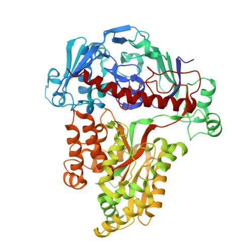
8A42, 8QLC, 8QLG, 8QLH, 8QLJ, 8QLK, 8QLM, 8QLV, 8QM0 - PubMed Abstract:
ATP-binding cassette (ABC) transport systems are crucial for bacteria to ensure sufficient uptake of nutrients that are not produced de novo or improve the energy balance. The cell surface of the pathobiont Streptococcus pneumoniae (pneumococcus) is decorated with a substantial array of ABC transporters, critically influencing nasopharyngeal colonization and invasive infections. Given the auxotrophic nature of pneumococci for certain amino acids, the Ami ABC transporter system, orchestrating oligopeptide uptake, becomes indispensable in host compartments lacking amino acids. The system comprises five exposed Oligopeptide Binding Proteins (OBPs) and four proteins building the ABC transporter channel. Here, we present a structural analysis of all the OBPs in this system. Multiple crystallographic structures, capturing both open and closed conformations along with complexes involving chemically synthesized peptides, have been solved at high resolution providing insights into the molecular basis of their diverse peptide specificities. Mass spectrometry analysis of oligopeptides demonstrates the unexpected remarkable promiscuity of some of these proteins when expressed in Escherichia coli, displaying affinity for a wide range of peptides. Finally, a model is proposed for the complete Ami transport system in complex with its various OBPs. We further disclosed, through in silico modelling, some essential structural changes facilitating oligopeptide transport into the cellular cytoplasm. Thus, the structural analysis of the Ami system provides valuable insights into the mechanism and specificity of oligopeptide binding by the different OBPs, shedding light on the intricacies of the uptake mechanism and the in vivo implications for this human pathogen.
- Department of Crystallography and Structural Biology, Institute of Physical-Chemistry "Blas Cabrera", Spanish National Research Council (CSIC), Madrid; Spain.
Organizational Affiliation: