Molecular Basis of Pseudomonas syringae pv actinidiae Levansucrase Inhibition by a Multivalent Iminosugar.
Cicchi, C., Pazzagli, L., Paoli, P., Campigli, S., Marchi, G., Cardona, F., Clemente, F., Pavone, S., Ferraroni, M., Canovai, A., Matassini, C., Luti, S.(2025) J Agric Food Chem 73: 15981-15992
- PubMed: 40349214 Search on PubMedSearch on PubMed Central
- DOI: https://doi.org/10.1021/acs.jafc.5c01947
- Primary Citation Related Structures:
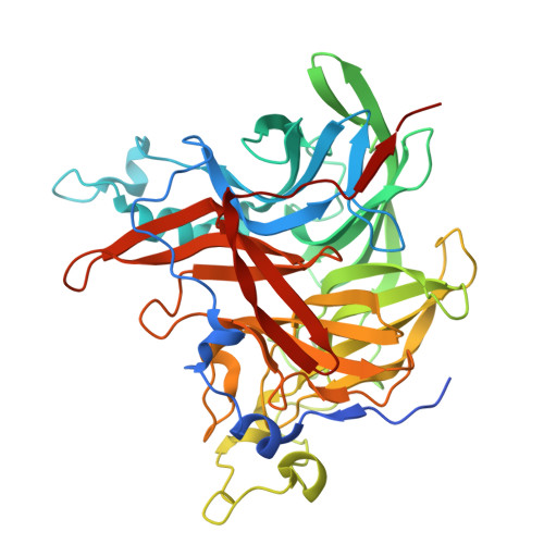
8QJ5, 8QKW - PubMed Abstract:
Levansucrases are a class of polysaccharide-processing enzymes widely distributed among plant pathogenic bacteria, such as Pseudomonas syringae and Erwinia amylovora . Therefore, the modulation of levansucrase activity could represent a new strategy to reduce the microbial survival of such bacteria. Herein, we identified a tetravalent pyrrolidine iminosugar (TPIS) as the first levansucrase inhibitor described to date. TPIS reversibly inhibits sucrose hydrolysis and levan polymerization of levansucrase derived from different bacterial genotypes of P. syringae , showing competitive behavior and an inhibition constant ( K i ) in the micromolar range. Interestingly, the monovalent pyrrolidine iminosugar (PIS) analogue shows negligible inhibition, suggesting that multivalency plays a pivotal role in the interaction with levansucrase. To gain insight into the binding mechanism, the X-ray crystal structures of the beta levansucrase isoform from P. syringae pv actinidiae (Psa) in its native form and in complex with TPIS were solved, confirming TPIS as a competitive inhibitor of levansucrases. Only a portion of TPIS, corresponding to one chain of the tetravalent iminosugar derivative, was visible in the electron density maps. Nevertheless, our structural data provided an adequate comprehension of the inhibitor/enzyme interactions, sufficient to exclude some of the possible inhibition mechanisms justifying a multivalent effect and pave the way for the development of new, more potent inhibitors.
- Department of Experimental and Clinical Biomedical Sciences, University of Florence, Viale Morgagni n. 50, Florence 50134, Italy.
Organizational Affiliation: