8QHR

Crystal structure of the human DNPH1 glycosyl-enzyme intermediate

  • Classification: HYDROLASE
  • Organism(s): Homo sapiens
  • Expression System: Escherichia coli
  • Mutation(s): Yes 

  • Deposited: 2023-09-09 Released: 2023-11-08 
  • Deposition Author(s): Rzechorzek, N.J., West, S.C.
  • Funding Organization(s): The Francis Crick Institute, Cancer Research UK, Medical Research Council (MRC, United Kingdom), Wellcome Trust, European Research Council (ERC), Louis-Jeantet Foundation

Experimental Data Snapshot

  • Method: X-RAY DIFFRACTION
  • Resolution: 1.65 Å
  • R-Value Free: 0.222 
  • R-Value Work: 0.184 
  • R-Value Observed: 0.186 

wwPDB Validation   3D Report Full Report


Ligand Structure Quality Assessment 


This is version 1.0 of the entry. See complete history


Literature

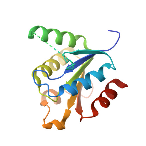
Mechanism of substrate hydrolysis by the human nucleotide pool sanitiser DNPH1.

Rzechorzek, N.J.Kunzelmann, S.Purkiss, A.G.Silva Dos Santos, M.MacRae, J.I.Taylor, I.A.Fugger, K.West, S.C.

(2023) Nat Commun 14: 6809-6809

  • DOI: https://doi.org/10.1038/s41467-023-42544-4
  • Primary Citation of Related Structures:  
    8QHQ, 8QHR

  • PubMed Abstract: 

    Poly(ADP-ribose) polymerase (PARP) inhibitors are used in the clinic to treat BRCA-deficient breast, ovarian and prostate cancers. As their efficacy is potentiated by loss of the nucleotide salvage factor DNPH1 there is considerable interest in the development of highly specific small molecule DNPH1 inhibitors. Here, we present X-ray crystal structures of dimeric DNPH1 bound to its substrate hydroxymethyl deoxyuridine monophosphate (hmdUMP). Direct interaction with the hydroxymethyl group is important for substrate positioning, while conserved residues surrounding the base facilitate target discrimination. Glycosidic bond cleavage is driven by a conserved catalytic triad and proceeds via a two-step mechanism involving formation and subsequent disruption of a covalent glycosyl-enzyme intermediate. Mutation of a previously uncharacterised yet conserved glutamate traps the intermediate in the active site, demonstrating its role in the hydrolytic step. These observations define the enzyme's catalytic site and mechanism of hydrolysis, and provide important insights for inhibitor discovery.


  • Organizational Affiliation

    DNA Recombination and Repair Laboratory, The Francis Crick Institute, 1 Midland Road, London, NW1 1AT, UK.


Macromolecules
Find similar proteins by:  (by identity cutoff)  |  3D Structure
Entity ID: 1
MoleculeChains Sequence LengthOrganismDetailsImage
2'-deoxynucleoside 5'-phosphate N-hydrolase 1
A, B
147Homo sapiensMutation(s): 1 
Gene Names: DNPH1C6orf108RCL
EC: 3.2.2
UniProt & NIH Common Fund Data Resources
Find proteins for O43598 (Homo sapiens)
Explore O43598 
Go to UniProtKB:  O43598
PHAROS:  O43598
GTEx:  ENSG00000112667 
Entity Groups  
Sequence Clusters30% Identity50% Identity70% Identity90% Identity95% Identity100% Identity
UniProt GroupO43598
Sequence Annotations
Expand
  • Reference Sequence
Experimental Data & Validation

Experimental Data

  • Method: X-RAY DIFFRACTION
  • Resolution: 1.65 Å
  • R-Value Free: 0.222 
  • R-Value Work: 0.184 
  • R-Value Observed: 0.186 
  • Space Group: P 21 21 21
Unit Cell:
Length ( Å )Angle ( ˚ )
a = 47.42α = 90
b = 55.039β = 90
c = 103.949γ = 90
Software Package:
Software NamePurpose
PHENIXrefinement
DIALSdata reduction
pointlessdata scaling
Aimlessdata scaling
PHASERphasing

Structure Validation

View Full Validation Report


Ligand Structure Quality Assessment 


Entry History & Funding Information

Deposition Data


Funding OrganizationLocationGrant Number
The Francis Crick InstituteUnited Kingdom--
Cancer Research UKUnited KingdomCC2098
Medical Research Council (MRC, United Kingdom)United KingdomCC2098
Wellcome TrustUnited KingdomCC2098
European Research Council (ERC)European UnionERC-ADG-666400
Louis-Jeantet FoundationSwitzerland--

Revision History  (Full details and data files)

  • Version 1.0: 2023-11-08
    Type: Initial release