8Q9Q

Crystal Structure of the MADS-box/MEF2 Domain of MEF2D bound to dsDNA and HDAC7 deacetylase binding motif


Experimental Data Snapshot

  • Method: X-RAY DIFFRACTION
  • Resolution: 2.11 Å
  • R-Value Free: 0.285 
  • R-Value Work: 0.265 
  • R-Value Observed: 0.266 

wwPDB Validation   3D Report Full Report


This is version 1.0 of the entry. See complete history


Literature

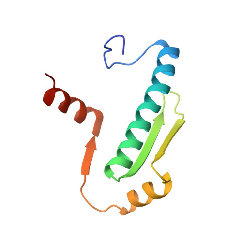
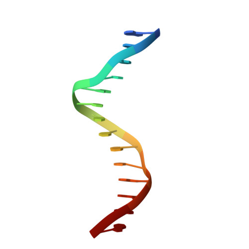
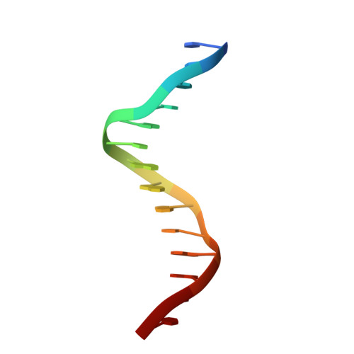
Folding of Class IIa HDAC Derived Peptides into alpha-helices Upon Binding to Myocyte Enhancer Factor-2 in Complex with DNA.

Chinellato, M.Perin, S.Carli, A.Lastella, L.Biondi, B.Borsato, G.Di Giorgio, E.Brancolini, C.Cendron, L.Angelini, A.

(2024) J Mol Biol 436: 168541-168541

  • DOI: https://doi.org/10.1016/j.jmb.2024.168541
  • Primary Citation of Related Structures:  
    8C84, 8PDE, 8Q9P, 8Q9Q, 8Q9R

  • PubMed Abstract: 

    Interaction of transcription factor myocyte enhancer factor-2 (MEF2) family members with class IIa histone deacetylases (HDACs) has been implicated in a wide variety of diseases. Though considerable knowledge on this topic has been accumulated over the years, a high resolution and detailed analysis of the binding mode of multiple class IIa HDAC derived peptides with MEF2D is still lacking. To fulfil this gap, we report here the crystal structure of MEF2D in complex with double strand DNA and four different class IIa HDAC derived peptides, namely HDAC4, HDAC5, HDAC7 and HDAC9. All class IIa HDAC derived peptides form extended amphipathic α-helix structures that fit snugly in the hydrophobic groove of MEF2D domain. Binding mode of class IIa HDAC derived peptides to MEF2D is very similar and occur primarily through nonpolar interactions mediated by highly conserved branched hydrophobic amino acids. Further studies revealed that class IIa HDAC derived peptides are unstructured in solution and appear to adopt a folded α-helix structure only upon binding to MEF2D. Comparison of our peptide-protein complexes with previously characterized structures of MEF2 bound to different co-activators and co-repressors, highlighted both differences and similarities, and revealed the adaptability of MEF2 in protein-protein interactions. The elucidation of the three-dimensional structure of MEF2D in complex with multiple class IIa HDAC derived peptides provide not only a better understanding of the molecular basis of their interactions but also have implications for the development of novel antagonist.


  • Organizational Affiliation

    Department of Biology, University of Padua, Via U. Bassi 58, 35131 Padova, Italy.


Macromolecules

Find similar proteins by:  Sequence   |   3D Structure  

Entity ID: 1
MoleculeChains Sequence LengthOrganismDetailsImage
HDAC7 (histone deacetylase 7) binding motif peptide: GLY-VAL-VAL-LYS-GLN-LYS-LEU-ALA-GLU-VAL-ILE-LEU-LYS-LYS-GLNA [auth X]15Homo sapiensMutation(s): 0 
Entity Groups  
Sequence Clusters30% Identity50% Identity70% Identity90% Identity95% Identity100% Identity
Sequence Annotations
Expand
  • Reference Sequence
Find similar proteins by:  (by identity cutoff)  |  3D Structure
Entity ID: 2
MoleculeChains Sequence LengthOrganismDetailsImage
MEF2D proteinB [auth A],
C [auth B]
95Homo sapiensMutation(s): 0 
Gene Names: MEF2D
UniProt & NIH Common Fund Data Resources
Find proteins for Q05BX2 (Homo sapiens)
Explore Q05BX2 
Go to UniProtKB:  Q05BX2
GTEx:  ENSG00000116604 
Entity Groups  
Sequence Clusters30% Identity50% Identity70% Identity90% Identity95% Identity100% Identity
UniProt GroupQ05BX2
Sequence Annotations
Expand
  • Reference Sequence

Find similar nucleic acids by:  Sequence   |   3D Structure  

Entity ID: 3
MoleculeChains LengthOrganismImage
MADS box dsDNA: AACTATTTATAAGAD [auth K]14DNA molecule
Sequence Annotations
Expand
  • Reference Sequence

Find similar nucleic acids by:  Sequence   |   3D Structure  

Entity ID: 4
MoleculeChains LengthOrganismImage
MADS box dsDNA: TCTTATAAATAGTTE [auth L]14DNA molecule
Sequence Annotations
Expand
  • Reference Sequence
Experimental Data & Validation

Experimental Data

  • Method: X-RAY DIFFRACTION
  • Resolution: 2.11 Å
  • R-Value Free: 0.285 
  • R-Value Work: 0.265 
  • R-Value Observed: 0.266 
  • Space Group: P 21 21 21
Unit Cell:
Length ( Å )Angle ( ˚ )
a = 56.883α = 90
b = 59.361β = 90
c = 76.502γ = 90
Software Package:
Software NamePurpose
REFMACrefinement
autoPROCdata reduction
Aimlessdata scaling
MOLREPphasing

Structure Validation

View Full Validation Report


Entry History & Funding Information

Deposition Data


Funding OrganizationLocationGrant Number
Italian Ministry of EducationItalyC94I19001550001

Revision History  (Full details and data files)

  • Version 1.0: 2024-04-17
    Type: Initial release