8Q8P | pdb_00008q8p

Cryo-EM structure of the magnesium channel CtMrs2 in the closed state


Experimental Data Snapshot

  • Method: ELECTRON MICROSCOPY
  • Resolution: 2.68 Å
  • Aggregation State: PARTICLE 
  • Reconstruction Method: SINGLE PARTICLE 

wwPDB Validation 3D Report Full Report

Validation slider image for 8Q8P

This is version 1.4 of the entry. See complete history

Literature

Closed and open structures of the eukaryotic magnesium channel Mrs2 reveal the auto-ligand-gating regulation mechanism.

Li, P.Liu, S.Wallerstein, J.Villones, R.L.E.Huang, P.Lindkvist-Petersson, K.Meloni, G.Lu, K.Steen Jensen, K.Liin, S.I.Gourdon, P.

(2025) Nat Struct Mol Biol 32: 491-501

  • DOI: https://doi.org/10.1038/s41594-024-01432-1
  • Primary Citation Related Structures: 
    8Q8P, 8Q8Q

  • PubMed Abstract: 

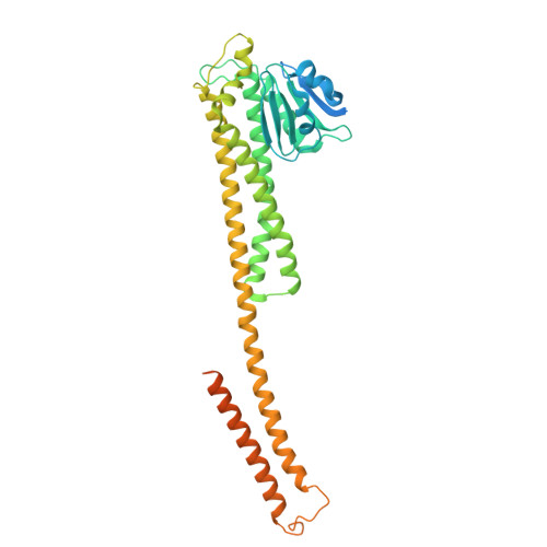
    The CorA/Mrs2 family of pentameric proteins are cardinal for the influx of Mg 2+ across cellular membranes, importing the cation to mitochondria in eukaryotes. Yet, the conducting and regulation mechanisms of permeation remain elusive, particularly for the eukaryotic Mrs2 members. Here, we report closed and open Mrs2 cryo-electron microscopy structures, accompanied by functional characterization. Mg 2+ flux is permitted by a narrow pore, gated by methionine and arginine residues in the closed state. Transition between the conformations is orchestrated by two pairs of conserved sensor-serving Mg 2+ -binding sites in the mitochondrial matrix lumen, located in between monomers. At lower levels of Mg 2+ , these ions are stripped, permitting an alternative, symmetrical shape, maintained by the RDLR motif that replaces one of the sensor site pairs in the open conformation. Thus, our findings collectively establish the molecular basis for selective Mg 2+ influx of Mrs2 and an auto-ligand-gating regulation mechanism.


  • Organizational Affiliation
    • Department of Experimental Medical Science, Lund University, Lund, Sweden. ping.li@med.lu.se.

Macromolecule Content 

  • Total Structure Weight: 241.98 kDa 
  • Atom Count: 12,579 
  • Modeled Residue Count: 1,585 
  • Deposited Residue Count: 2,125 
  • Unique protein chains: 1

Macromolecules

Find similar proteins by:|  3D Structure
Entity ID: 1
MoleculeChains  Sequence LengthOrganismDetailsImage
Magnesium channel Mrs2
A, B, C, D, E
425Thermochaetoides thermophilaMutation(s): 0 
Membrane Entity: Yes 
UniProt
Find proteins for G0S186 (Chaetomium thermophilum (strain DSM 1495 / CBS 144.50 / IMI 039719))
Explore G0S186 
Go to UniProtKB:  G0S186
Entity Groups
Sequence Clusters30% Identity50% Identity70% Identity90% Identity95% Identity100% Identity
UniProt GroupG0S186
Sequence Annotations
Expand
Reference Sequence

Small Molecules

Ligands 1 Unique
IDChains Name / Formula / InChI Key2D Diagram3D Interactions
MG
(Subject of Investigation/LOI)

Query on MG


Download:Ideal Coordinates CCD File
AA [auth E]
BA [auth E]
CA [auth E]
F [auth A]
G [auth A]
AA [auth E],
BA [auth E],
CA [auth E],
F [auth A],
G [auth A],
H [auth A],
I [auth A],
J [auth A],
K [auth A],
L [auth A],
M [auth A],
N [auth B],
O [auth B],
P [auth B],
Q [auth C],
R [auth C],
S [auth C],
T [auth C],
U [auth C],
V [auth D],
W [auth D],
X [auth D],
Y [auth D],
Z [auth E]
MAGNESIUM ION
Mg
JLVVSXFLKOJNIY-UHFFFAOYSA-N

Experimental Data & Validation

Experimental Data

  • Method: ELECTRON MICROSCOPY
  • Resolution: 2.68 Å
  • Aggregation State: PARTICLE 
  • Reconstruction Method: SINGLE PARTICLE 

Structure Validation

View Full Validation Report


Entry History 

& Funding Information

Deposition Data


Funding OrganizationLocationGrant Number
Swedish Research CouncilSweden--

Revision History  (Full details and data files)

  • Version 1.0: 2024-11-27
    Type: Initial release
  • Version 1.1: 2024-12-04
    Changes: Data collection, Database references
  • Version 1.2: 2024-12-11
    Changes: Data collection, Database references
  • Version 1.3: 2024-12-25
    Changes: Data collection, Database references, Structure summary
  • Version 1.4: 2025-03-26
    Changes: Data collection, Database references