Structure-Based Design of a Potent and Selective YTHDC1 Ligand.
Zalesak, F., Nai, F., Herok, M., Bochenkova, E., Bedi, R.K., Li, Y., Errani, F., Caflisch, A.(2024) J Med Chem 67: 9516-9535
- PubMed: 38787793 Search on PubMed
- DOI: https://doi.org/10.1021/acs.jmedchem.4c00599
- Primary Citation Related Structures:
8Q2Q, 8Q2R, 8Q2S, 8Q2T, 8Q2U, 8Q2V, 8Q2W, 8Q2X, 8Q2Y, 8Q31, 8Q32, 8Q33, 8Q35, 8Q37, 8Q38, 8Q39, 8Q3A, 8Q3G, 8Q4M, 8Q4N, 8Q4P, 8Q4Q, 8Q4R, 8Q4T, 8Q4U, 8Q4V, 8Q4W - PubMed Abstract:
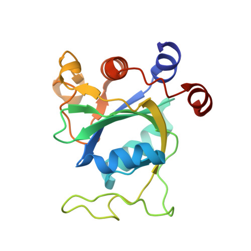
N 6 -Adenosine methylation (m 6 A) is a prevalent post-transcriptional modification of mRNA, with YTHDC1 being the reader protein responsible for recognizing this modification in the cell nucleus. Here, we present a protein structure-based medicinal chemistry campaign that resulted in the YTHDC1 inhibitor 40 , which shows an equilibrium dissociation constant ( K d ) of 49 nM. The crystal structure of the complex (1.6 Å resolution) validated the design. Compound 40 is selective against the cytoplasmic m 6 A-RNA readers YTHDF1-3 and YTHDC2 and shows antiproliferative activity against the acute myeloid leukemia (AML) cell lines THP-1, MOLM-13, and NOMO-1. For the series of compounds that culminated into ligand 40 , the good correlation between the affinity in the biochemical assay and antiproliferative activity in the THP-1 cell line provides evidence of YTHDC1 target engagement in the cell. The binding to YTHDC1 in the cell is further supported by the cellular thermal shift assay. Thus, ligand 40 is a tool compound for studying the role of YTHDC1 in AML.
- Department of Biochemistry, University of Zurich, Winterthurerstrasse 190, CH-8057 Zurich, Switzerland.
Organizational Affiliation: