8Q2C | pdb_00008q2c

Crystal structure of the E. coli PqiC Lipoprotein


Experimental Data Snapshot

  • Method: X-RAY DIFFRACTION
  • Resolution: 3.21 Å
  • R-Value Free: 
    0.306 (Depositor), 0.303 (DCC) 
  • R-Value Work: 
    0.251 (Depositor), 0.250 (DCC) 
  • R-Value Observed: 
    0.253 (Depositor) 

Starting Model: in silico
View more details

wwPDB Validation 3D Report Full Report

Validation slider image for 8Q2C

This is version 1.1 of the entry. See complete history

Literature

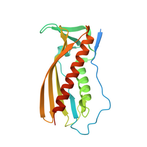
An octameric PqiC toroid stabilises the outer-membrane interaction of the PqiABC transport system.

Cooper, B.F.Ratkeviciute, G.Clifton, L.A.Johnston, H.Holyfield, R.Hardy, D.J.Caulton, S.G.Chatterton, W.Sridhar, P.Wotherspoon, P.Hughes, G.W.Hall, S.C.Lovering, A.L.Knowles, T.J.

(2024) EMBO Rep 25: 82-101

  • DOI: https://doi.org/10.1038/s44319-023-00014-4
  • Primary Citation Related Structures: 
    8Q2C, 8Q2D

  • PubMed Abstract: 

    The E. coli Paraquat Inducible (Pqi) Pathway is a putative Gram-negative phospholipid transport system. The pathway comprises three components: an integral inner membrane protein (PqiA), a periplasmic spanning MCE family protein (PqiB) and an outer membrane lipoprotein (PqiC). Interactions between all complex components, including stoichiometry, remain uncharacterised; nevertheless, once assembled into their quaternary complex, the trio of Pqi proteins are anticipated to provide a continuous channel between the inner and outer membranes of diderms. Here, we present X-ray structures of both the native and a truncated, soluble construct of the PqiC lipoprotein, providing insight into its biological assembly, and utilise neutron reflectometry to characterise the nature of the PqiB-PqiC-membrane interaction. Finally, we employ phenotypic complementation assays to probe specific PqiC residues, which imply the interaction between PqiB and PqiC is less intimate than previously anticipated.


  • Organizational Affiliation
    • Sir William Dunn School of Pathology, University of Oxford, OX1 3RE, Oxford, UK.

Macromolecule Content 

  • Total Structure Weight: 43.44 kDa 
  • Atom Count: 2,580 
  • Modeled Residue Count: 330 
  • Deposited Residue Count: 390 
  • Unique protein chains: 1

Macromolecules

Find similar proteins by:|  3D Structure
Entity ID: 1
MoleculeChains  Sequence LengthOrganismDetailsImage
Intermembrane transport lipoprotein PqiCA [auth B],
B [auth A]
195Escherichia coliMutation(s): 0 
Gene Names: pqiCymbAb0952JW5127
UniProt
Find proteins for P0AB10 (Escherichia coli (strain K12))
Explore P0AB10 
Go to UniProtKB:  P0AB10
Entity Groups
Sequence Clusters30% Identity50% Identity70% Identity90% Identity95% Identity100% Identity
UniProt GroupP0AB10
Sequence Annotations
Expand
Reference Sequence

Experimental Data & Validation

Experimental Data

  • Method: X-RAY DIFFRACTION
  • Resolution: 3.21 Å
  • R-Value Free:  0.306 (Depositor), 0.303 (DCC) 
  • R-Value Work:  0.251 (Depositor), 0.250 (DCC) 
  • R-Value Observed: 0.253 (Depositor) 
Space Group: I 4 2 2
Unit Cell:
Length ( Å )Angle ( ˚ )
a = 132.148α = 90
b = 132.148β = 90
c = 185.043γ = 90
Software Package:
Software NamePurpose
PHENIXrefinement
autoPROCdata reduction
STARANISOdata scaling
PHASERphasing

Structure Validation

View Full Validation Report


Entry History 

& Funding Information

Deposition Data


Funding OrganizationLocationGrant Number
Biotechnology and Biological Sciences Research Council (BBSRC)United KingdomBB/M01116X/1
Biotechnology and Biological Sciences Research Council (BBSRC)United KingdomBB/S017283/1

Revision History  (Full details and data files)

  • Version 1.0: 2023-09-20
    Type: Initial release
  • Version 1.1: 2024-01-31
    Changes: Database references