8PZQ | pdb_00008pzq

Model for focused reconstruction of influenza A RNP-like particle


Experimental Data Snapshot

  • Method: ELECTRON MICROSCOPY
  • Resolution: 5.30 Å
  • Aggregation State: FILAMENT 
  • Reconstruction Method: HELICAL 

Starting Model: experimental
View more details

wwPDB Validation 3D Report Full Report

Validation slider image for 8PZQ

This is version 1.0 of the entry. See complete history

Literature

Cryo-EM structure of influenza helical nucleocapsid reveals NP-NP and NP-RNA interactions as a model for the genome encapsidation.

Chenavier, F.Estrozi, L.F.Teulon, J.M.Zarkadas, E.Freslon, L.L.Pellequer, J.L.Ruigrok, R.W.H.Schoehn, G.Ballandras-Colas, A.Crepin, T.

(2023) Sci Adv 9: eadj9974-eadj9974

  • DOI: https://doi.org/10.1126/sciadv.adj9974
  • Primary Citation Related Structures: 
    8PZP, 8PZQ

  • PubMed Abstract: 

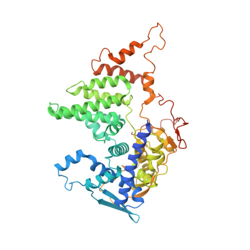
    Influenza virus genome encapsidation is essential for the formation of a helical viral ribonucleoprotein (vRNP) complex composed of nucleoproteins (NP), the trimeric polymerase, and the viral genome. Although low-resolution vRNP structures are available, it remains unclear how the viral RNA is encapsidated and how NPs assemble into the helical filament specific of influenza vRNPs. In this study, we established a biological tool, the RNP-like particles assembled from recombinant influenza A virus NP and synthetic RNA, and we present the first subnanometric cryo-electron microscopy structure of the helical NP-RNA complex (8.7 to 5.3 Å). The helical RNP-like structure reveals a parallel double-stranded conformation, allowing the visualization of NP-NP and NP-RNA interactions. The RNA, located at the interface of neighboring NP protomers, interacts with conserved residues previously described as essential for the NP-RNA interaction. The NP undergoes conformational changes to enable RNA binding and helix formation. Together, our findings provide relevant insights for understanding the mechanism for influenza genome encapsidation.


  • Organizational Affiliation
    • Univ. Grenoble Alpes, CNRS, CEA, IBS, F-38000, Grenoble, France.

Macromolecule Content 

  • Total Structure Weight: 179.82 kDa 
  • Atom Count: 11,420 
  • Modeled Residue Count: 1,432 
  • Deposited Residue Count: 1,542 
  • Unique protein chains: 1
  • Unique nucleic acid chains: 1

Macromolecules


Find similar proteins by:|  3D Structure
Entity ID: 1
MoleculeChains  Sequence LengthOrganismDetailsImage
NucleoproteinA,
D [auth B],
E [auth C]
506Influenza A virusMutation(s): 0 
Gene Names: NP
UniProt
Find proteins for A0ABF7SXN4 (Influenza A virus)
Explore A0ABF7SXN4 
Go to UniProtKB:  A0ABF7SXN4
Entity Groups
Sequence Clusters30% Identity50% Identity70% Identity90% Identity95% Identity100% Identity
UniProt GroupA0ABF7SXN4
Sequence Annotations
Expand
Reference Sequence
Find similar nucleic acids by:  Sequence
Entity ID: 2
MoleculeChains LengthOrganismImage
RNA (5'P-(UC)6-FAM3')B [auth D],
C [auth E]
12synthetic construct
Sequence Annotations
Expand
Reference Sequence

Experimental Data & Validation

Experimental Data

  • Method: ELECTRON MICROSCOPY
  • Resolution: 5.30 Å
  • Aggregation State: FILAMENT 
  • Reconstruction Method: HELICAL 
EM Software:
TaskSoftware PackageVersion
MODEL REFINEMENTPHENIX1.20.1_4487:
MODEL REFINEMENTCoot0.9.6
RECONSTRUCTIONcryoSPARC3.3.2

Structure Validation

View Full Validation Report


Entry History 

& Funding Information

Deposition Data


Funding OrganizationLocationGrant Number
Grenoble Alliance for Integrated Structural Cell Biology (GRAL)FranceANR-17-EURE-0003

Revision History  (Full details and data files)

  • Version 1.0: 2023-12-27
    Type: Initial release