8PZ7

crystal structure of VDR complex with D-Bishomo-1a,25-dihydroxyvitamin D3 Analog 57


Experimental Data Snapshot

  • Method: X-RAY DIFFRACTION
  • Resolution: 2.93 Å
  • R-Value Free: 0.257 
  • R-Value Work: 0.196 
  • R-Value Observed: 0.202 

wwPDB Validation   3D Report Full Report


Ligand Structure Quality Assessment 


This is version 1.1 of the entry. See complete history


Literature

Design, synthesis, and biological activity of D-bishomo-1 alpha ,25-dihydroxyvitamin D 3 analogs and their crystal structures with the vitamin D nuclear receptor.

Fabisiak, A.Brzeminski, P.Sicinski, R.R.Rochel, N.Maj, E.Filip-Psurska, B.Wietrzyk, J.Plum, L.A.DeLuca, H.F.

(2024) Eur J Med Chem 271: 116403-116403

  • DOI: https://doi.org/10.1016/j.ejmech.2024.116403
  • Primary Citation of Related Structures:  
    8PZ6, 8PZ7, 8PZ8, 8PZ9, 8PZB

  • PubMed Abstract: 

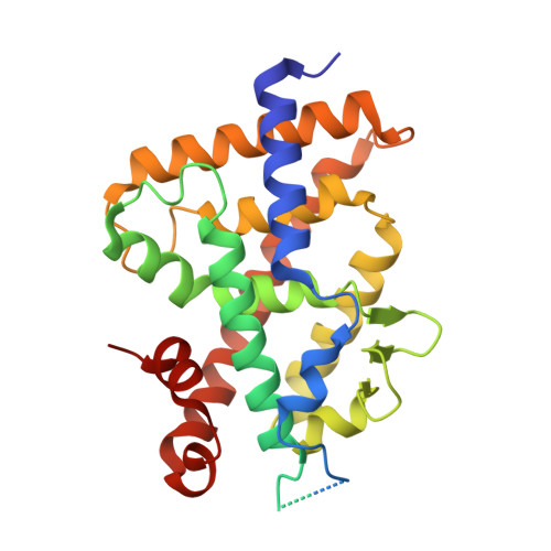
    The biologically active metabolite of vitamin D 3 - calcitriol - is a hormone involved in the regulation of calcium-phosphate homeostasis, immunological processes and cell differentiation, being therefore essential for the proper functioning of the human body. This suggests many applications of this steroid in the treatment of diseases such as rickets, psoriasis and some cancers. Unfortunately, using therapeutic doses of calcitriol is associated with high concentrations of this compound which causes hypercalcemia. For this reason, new calcitriol analogs are constantly sought, devoid of calcemic effects but maintaining its beneficial properties. In this study, we present the synthesis of vitamin D derivatives characterized by an enlarged (seven-membered) ring D. Preparation of the designed vitamin D compounds required separate syntheses of crucial building blocks (C/D-rings fragments with side chain and rings A) which were combined by different methods, including Wittig-Horner reaction and Suzuki coupling. Biological activities of the target vitamin D analogs were assessed both in vitro and in vivo, demonstrating their significant potency compared to the natural hormone. Furthermore, the successful crystallization of these compounds with the vitamin D receptor (VDR) enabled us to investigate additional molecular interactions with this protein.


  • Organizational Affiliation

    Department of Chemistry, University of Warsaw, Pasteura 1, 02-093, Warsaw, Poland. Electronic address: afabisiak@chem.uw.edu.pl.


Macromolecules
Find similar proteins by:  (by identity cutoff)  |  3D Structure
Entity ID: 1
MoleculeChains Sequence LengthOrganismDetailsImage
Vitamin D3 receptor A302Danio rerioMutation(s): 0 
Gene Names: vdranr1i1avdr
UniProt
Find proteins for Q9PTN2 (Danio rerio)
Explore Q9PTN2 
Go to UniProtKB:  Q9PTN2
Entity Groups  
Sequence Clusters30% Identity50% Identity70% Identity90% Identity95% Identity100% Identity
UniProt GroupQ9PTN2
Sequence Annotations
Expand
  • Reference Sequence

Find similar proteins by:  Sequence   |   3D Structure  

Entity ID: 2
MoleculeChains Sequence LengthOrganismDetailsImage
Nuclear receptor coactivator 213Homo sapiensMutation(s): 0 
UniProt & NIH Common Fund Data Resources
Find proteins for Q15596 (Homo sapiens)
Explore Q15596 
Go to UniProtKB:  Q15596
PHAROS:  Q15596
GTEx:  ENSG00000140396 
Entity Groups  
Sequence Clusters30% Identity50% Identity70% Identity90% Identity95% Identity100% Identity
UniProt GroupQ15596
Sequence Annotations
Expand
  • Reference Sequence
Small Molecules
Ligands 1 Unique
IDChains Name / Formula / InChI Key2D Diagram3D Interactions
IG0 (Subject of Investigation/LOI)
Query on IG0

Download Ideal Coordinates CCD File 
C [auth A](1~{R},3~{R})-5-[(2~{E})-2-[(4~{a}~{R},5~{R},9~{a}~{S})-4~{a}-methyl-5-[(2~{R})-6-methyl-6-oxidanyl-heptan-2-yl]-3,4,5,8,9,9~{a}-hexahydro-2~{H}-benzo[7]annulen-1-ylidene]ethylidene]-2-methyl-cyclohexane-1,3-diol
C29 H50 O3
AVNUSPMNPZNONM-DHXJDUGVSA-N
Experimental Data & Validation

Experimental Data

  • Method: X-RAY DIFFRACTION
  • Resolution: 2.93 Å
  • R-Value Free: 0.257 
  • R-Value Work: 0.196 
  • R-Value Observed: 0.202 
  • Space Group: P 65 2 2
Unit Cell:
Length ( Å )Angle ( ˚ )
a = 66.24α = 90
b = 66.24β = 90
c = 264.04γ = 120
Software Package:
Software NamePurpose
PHENIXrefinement
XDSdata reduction
Aimlessdata scaling
PHENIXphasing

Structure Validation

View Full Validation Report


Ligand Structure Quality Assessment 


Entry History & Funding Information

Deposition Data

  • Released Date: 2023-08-30 
  • Deposition Author(s): Rochel, N.

Funding OrganizationLocationGrant Number
Agence Nationale de la Recherche (ANR)FranceANR-21-CE17-0009-01

Revision History  (Full details and data files)

  • Version 1.0: 2023-08-30
    Type: Initial release
  • Version 1.1: 2024-04-24
    Changes: Database references