8PWM | pdb_00008pwm

Crystal structure of VDR in complex with Des-C-Ring and Aromatic-D-Ring analog 3b


Experimental Data Snapshot

  • Method: X-RAY DIFFRACTION
  • Resolution: 2.30 Å
  • R-Value Free: 
    0.252 (Depositor), 0.260 (DCC) 
  • R-Value Work: 
    0.196 (Depositor), 0.210 (DCC) 
  • R-Value Observed: 
    0.201 (Depositor) 

Starting Model: experimental
View more details

wwPDB Validation   3D Report Full Report


Ligand Structure Quality Assessment 


This is version 1.1 of the entry. See complete history


Literature

Further Studies on the Highly Active Des-C-Ring and Aromatic-D-Ring Analogues of 1 alpha ,25-Dihydroxyvitamin D 3 (Calcitriol): Refinement of the Side Chain.

Zarate-Ruiz, A.Seoane, S.Peluso-Iltis, C.Peters, S.Gregorio, C.Guiberteau, T.Maestro, M.Perez-Fernandez, R.Rochel, N.Mourino, A.

(2023) J Med Chem 66: 15326-15339

  • DOI: https://doi.org/10.1021/acs.jmedchem.3c01371
  • Primary Citation of Related Structures:  
    8PWD, 8PWE, 8PWF, 8PWM

  • PubMed Abstract: 

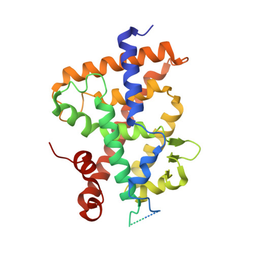
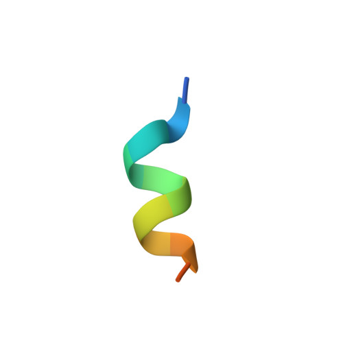
    Current efforts in the vitamin D field are directed toward the development of highly antiproliferative yet noncalcemic analogues of the natural hormone 1α,25-dihydroxyvitamin D 3 (1,25D 3 ). We have recently reported the design, synthesis, biological evaluation, and crystal structures of a series of novel analogues that both lack the steroidal C-ring and have an m -phenylene ring replacing the steroidal cyclopentane D-ring. We have now investigated the potentiating effects of incorporating selected modifications (hexafluorination and/or an internal triple bond) within the steroidal side chain in our series. An alternative synthetic strategy (Wittig-Horner approach instead of our previously used Pd-catalyzed tandem cyclization/cross-coupling) for the construction of the vitamin D triene system was found convenient for the target compounds 2 , 3a , 3b , and 3c of this report. These modifications enhance vitamin D nuclear receptor (VDR) interactions and consequently VDR-associated biological properties compared to parental PG-136 compound while maintaining normal calcium levels.


  • Organizational Affiliation
    • Department of Organic Chemistry, Research Laboratory Ignacio Ribas, University of Santiago de Compostela, Avda. de las Ciencias s/n, Santiago de Compostela 15782, Spain.

Macromolecules
Find similar proteins by:  (by identity cutoff)  |  3D Structure
Entity ID: 1
MoleculeChains Sequence LengthOrganismDetailsImage
Vitamin D3 receptor A302Danio rerioMutation(s): 0 
Gene Names: vdranr1i1avdr
UniProt
Find proteins for Q9PTN2 (Danio rerio)
Explore Q9PTN2 
Go to UniProtKB:  Q9PTN2
Entity Groups  
Sequence Clusters30% Identity50% Identity70% Identity90% Identity95% Identity100% Identity
UniProt GroupQ9PTN2
Sequence Annotations
Expand
  • Reference Sequence

Find similar proteins by:  Sequence   |   3D Structure  

Entity ID: 2
MoleculeChains Sequence LengthOrganismDetailsImage
Nuclear receptor coactivator 213Homo sapiensMutation(s): 0 
UniProt & NIH Common Fund Data Resources
Find proteins for Q15596 (Homo sapiens)
Explore Q15596 
Go to UniProtKB:  Q15596
PHAROS:  Q15596
GTEx:  ENSG00000140396 
Entity Groups  
Sequence Clusters30% Identity50% Identity70% Identity90% Identity95% Identity100% Identity
UniProt GroupQ15596
Sequence Annotations
Expand
  • Reference Sequence
Small Molecules
Ligands 2 Unique
IDChains Name / Formula / InChI Key2D Diagram3D Interactions
HXI (Subject of Investigation/LOI)
Query on HXI

Download Ideal Coordinates CCD File 
D [auth A](1R,3S,5Z)-4-methylidene-5-[(E)-3-[3-[7,7,7-tris(fluoranyl)-6-oxidanyl-6-(trifluoromethyl)hept-3-ynyl]phenyl]but-2-enylidene]cyclohexane-1,3-diol
C25 H26 F6 O3
YPFOJCHONFJOGI-SGTALLODSA-N
ACT
Query on ACT

Download Ideal Coordinates CCD File 
C [auth A]ACETATE ION
C2 H3 O2
QTBSBXVTEAMEQO-UHFFFAOYSA-M
Experimental Data & Validation

Experimental Data

  • Method: X-RAY DIFFRACTION
  • Resolution: 2.30 Å
  • R-Value Free:  0.252 (Depositor), 0.260 (DCC) 
  • R-Value Work:  0.196 (Depositor), 0.210 (DCC) 
  • R-Value Observed: 0.201 (Depositor) 
Space Group: P 65 2 2
Unit Cell:
Length ( Å )Angle ( ˚ )
a = 65.849α = 90
b = 65.849β = 90
c = 264.213γ = 120
Software Package:
Software NamePurpose
PHENIXrefinement
XDSdata reduction
Aimlessdata scaling
PHENIXphasing

Structure Validation

View Full Validation Report


Ligand Structure Quality Assessment 


Entry History & Funding Information

Deposition Data

  • Released Date: 2024-08-21 
  • Deposition Author(s): Rochel, N.

Funding OrganizationLocationGrant Number
Agence Nationale de la Recherche (ANR)France--

Revision History  (Full details and data files)

  • Version 1.0: 2024-08-21
    Type: Initial release
  • Version 1.1: 2025-03-12
    Changes: Database references, Structure summary