


Structure of the toxin/antitoxin complex FaRel/ATfaRel2 with APCPP
Garcia-Pino, A., Talavera Perez, A., Dominguez Molina, L.To be published.
Experimental Data Snapshot
Starting Model: experimental
View more details
Entity ID: 1 | |||||
|---|---|---|---|---|---|
| Molecule | Chains | Sequence Length | Organism | Details | Image |
| ATfaRel2 | 75 | Thomasclavelia ramosa | Mutation(s): 0 Gene Names: DXB93_19740 | 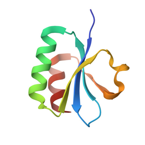 | |
UniProt | |||||
Entity Groups | |||||
| Sequence Clusters | 30% Identity50% Identity70% Identity90% Identity95% Identity100% Identity | ||||
| UniProt Group | A0A3E3DY87 | ||||
Sequence AnnotationsExpand | |||||
Reference Sequence | |||||
Entity ID: 2 | |||||
|---|---|---|---|---|---|
| Molecule | Chains | Sequence Length | Organism | Details | Image |
| RelA/SpoT domain-containing protein | 207 | Thomasclavelia ramosa | Mutation(s): 1 Gene Names: DXB93_19735 | 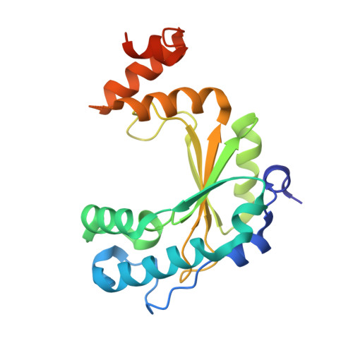 | |
UniProt | |||||
Entity Groups | |||||
| Sequence Clusters | 30% Identity50% Identity70% Identity90% Identity95% Identity100% Identity | ||||
| UniProt Group | A0A3E3DY42 | ||||
Sequence AnnotationsExpand | |||||
Reference Sequence | |||||
| Ligands 5 Unique | |||||
|---|---|---|---|---|---|
| ID | Chains | Name / Formula / InChI Key | 2D Diagram | 3D Interactions | |
| APC (Subject of Investigation/LOI) Download:Ideal Coordinates CCD File | P [auth B] | DIPHOSPHOMETHYLPHOSPHONIC ACID ADENOSYL ESTER C11 H18 N5 O12 P3 CAWZRIXWFRFUQB-IOSLPCCCSA-N |  | ||
| CAC Download:Ideal Coordinates CCD File | D [auth A] | CACODYLATE ION C2 H6 As O2 OGGXGZAMXPVRFZ-UHFFFAOYSA-M |  | ||
| GOL Download:Ideal Coordinates CCD File | F [auth A], Q [auth B], R [auth B] | GLYCEROL C3 H8 O3 PEDCQBHIVMGVHV-UHFFFAOYSA-N |  | ||
| POL Download:Ideal Coordinates CCD File | C [auth A] E [auth A] G [auth A] I [auth A] J [auth A] | N-PROPANOL C3 H8 O BDERNNFJNOPAEC-UHFFFAOYSA-N |  | ||
| MG Download:Ideal Coordinates CCD File | H [auth A], K [auth B], L [auth B] | MAGNESIUM ION Mg JLVVSXFLKOJNIY-UHFFFAOYSA-N |  | ||
| Length ( Å ) | Angle ( ˚ ) |
|---|---|
| a = 227.81 | α = 90 |
| b = 227.81 | β = 90 |
| c = 227.81 | γ = 90 |
| Software Name | Purpose |
|---|---|
| BUSTER | refinement |
| autoPROC | data reduction |
| XSCALE | data scaling |
| PHASER | phasing |
| Funding Organization | Location | Grant Number |
|---|---|---|
| Fonds National de la Recherche Scientifique (FNRS) | Belgium | PDR T.0090.22 |


