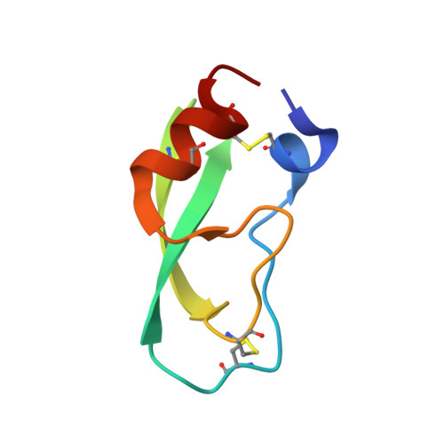
8PTI

CRYSTAL STRUCTURE OF A Y35G MUTANT OF BOVINE PANCREATIC TRYPSIN INHIBITOR


Experimental Data Snapshot

  • Method: X-RAY DIFFRACTION
  • Resolution: 1.80 Å
  • R-Value Observed: 0.159 

wwPDB Validation   3D Report Full Report


This is version 1.2 of the entry. See complete history


Literature

Crystal structure of a Y35G mutant of bovine pancreatic trypsin inhibitor.

Housset, D.Kim, K.S.Fuchs, J.Woodward, C.Wlodawer, A.

(1991) J Mol Biol 220: 757-770

  • DOI: https://doi.org/10.1016/0022-2836(91)90115-m
  • Primary Citation of Related Structures:  
    8PTI

  • PubMed Abstract: 
  • The structure of a Y35G mutant of bovine pancreatic trypsin inhibitor (BPTI) was solved by molecular replacement and was refined by both simulated annealing and restrained least-squares at 1.8 A resolution. The crystals belong to the space group P42212, with unit cell dimensions a = b = 46 ...

    The structure of a Y35G mutant of bovine pancreatic trypsin inhibitor (BPTI) was solved by molecular replacement and was refined by both simulated annealing and restrained least-squares at 1.8 A resolution. The crystals belong to the space group P42212, with unit cell dimensions a = b = 46.75 A, c = 50.61 A. The final R-factor is 0.159 and the deviation from ideality for bond distances is 0.02 A. The structure of the mutant differs from that of the native protein, showing an overall root-mean-square (r.m.s.) difference of 1.86 A for main-chain atoms. However, the change is mostly localized in the two loops (respective r.m.s. values of 2.04 A and 3.93 A) and the C terminus (r.m.s. 6.79 A), while the core of the protein is well conserved (r.m.s. 0.45 A). The change in the loop regions can be clearly attributed to the mutation while the difference in the C terminus might be only due to a different crystal packing. Seventy water molecules were included in the model but only seven of them are shared with the native structure. Thermal parameters are showing a good correlation with those for the wild-type of BPTI.


    Related Citations: 
    • Structural Effects Induced by Removal of a Disulfide-Bridge. The X-Ray Structure of the C30A(Slash)C51A Mutant of Basic Pancreatic Trypsin Inhibitor at 1.6 Angstroms
      Eigenbrot, C., Randal, M., Kossiakoff, A.A.
      (1990) Protein Eng 3: 591
    • X-Ray Crystal Structure of the Protease Inhibitor Domain of Alzheimer'S Amyloid Beta-Protein Precursor
      Hynes, T.R., Randal, M., Kennedy, L.A., Eigenbrot, C., Kossiakoff, A.A.
      (1990) Biochemistry 29: 10018
    • Structure of Form III Crystals of Bovine Pancreatic Trypsin Inhibitor.
      Wlodawer, A., Nachman, J., Gilliland, G.L., Gallagher, W., Woodward, C.
      (1987) J Mol Biol 198: 469
    • Structure of Bovine Pancreatic Trypsin Inhibitor. Results of Joint Neutron and X-Ray Refinement of Crystal Form II
      Wlodawer, A., Walter, J., Huber, R., Sjolin, L.
      (1984) J Mol Biol 180: 307
    • Comparison of Two Highly Refined Structures of Bovine Pancreatic Trypsin Inhibitor
      Wlodawer, A., Deisenhofer, J., Huber, R.
      (1987) J Mol Biol 193: 145
    • Pancreatic Trypsin Inhibitor. A New Crystal Form and its Analysis
      Walter, J., Huber, R.
      (1983) J Mol Biol 167: 911

    Organizational Affiliation

    Macromolecular Structure Laboratory, NCI-Frederick Cancer Research and Development Center, MD 21702.


Macromolecules
Find similar proteins by:  (by identity cutoff)  |  3D Structure
Entity ID: 1
MoleculeChainsSequence LengthOrganismDetailsImage
BOVINE PANCREATIC TRYPSIN INHIBITOR58Bos taurusMutation(s): 0 
UniProt
Find proteins for P00974 (Bos taurus)
Explore P00974 
Go to UniProtKB:  P00974
Entity Groups  
Sequence Clusters30% Identity50% Identity70% Identity90% Identity95% Identity100% Identity
UniProt GroupP00974
Protein Feature View
Expand
  • Reference Sequence
Experimental Data & Validation

Experimental Data

  • Method: X-RAY DIFFRACTION
  • Resolution: 1.80 Å
  • R-Value Observed: 0.159 
  • Space Group: P 42 21 2
Unit Cell:
Length ( Å )Angle ( ˚ )
a = 46.75α = 90
b = 46.75β = 90
c = 50.61γ = 90
Software Package:
Software NamePurpose
PROLSQrefinement

Structure Validation

View Full Validation Report


Entry History 

Deposition Data

Revision History  (Full details and data files)

  • Version 1.0: 1991-04-15
    Type: Initial release
  • Version 1.1: 2008-03-25
    Changes: Version format compliance
  • Version 1.2: 2011-07-13
    Changes: Version format compliance