Structural characterization of PHOX2B and its DNA interaction shed light on the molecular basis of the +7Ala variant pathogenicity in CCHS.
Diana, D., Pirone, L., Russo, L., D'Abrosca, G., Madheswaran, M., Benfante, R., Di Lascio, S., Caldinelli, L., Fornasari, D., Acconcia, C., Corvino, A., Ventserova, N., Pollegioni, L., Isernia, C., Di Gaetano, S., Malgieri, G., Pedone, E.M., Fattorusso, R.(2024) Chem Sci 15: 8858-8872
- PubMed: 38873078 Search on PubMedSearch on PubMed Central
- DOI: https://doi.org/10.1039/d3sc06427a
- Primary Citation Related Structures:
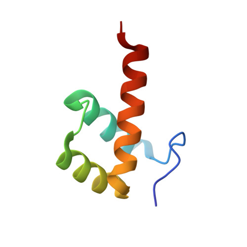
8P7G - PubMed Abstract:
An expansion of poly-alanine up to +13 residues in the C-terminus of the transcription factor PHOX2B underlies the onset of congenital central hypoventilation syndrome (CCHS). Recent studies demonstrated that the alanine tract expansion influences PHOX2B folding and activity. Therefore, structural information on PHOX2B is an important target for obtaining clues to elucidate the insurgence of the alanine expansion-related syndrome and also for defining a viable therapy. Here we report by NMR spectroscopy the structural characterization of the homeodomain (HD) of PHOX2B and HD + C-terminus PHOX2B protein, free and in the presence of the target DNA. The obtained structural data are then exploited to obtain a structural model of the PHOX2B-DNA interaction. In addition, the variant +7Ala, responsible for one of the most frequent forms of the syndrome, was analysed, showing different conformational proprieties in solution and a strong propensity to aggregation. Our data suggest that the elongated poly-alanine tract would be related to disease onset through a loss-of-function mechanism. Overall, this study paves the way for the future rational design of therapeutic drugs, suggesting as a possible therapeutic route the use of specific anti-aggregating molecules capable of preventing variant aggregation and possibly restoring the DNA-binding activity of PHOX2B.
- CNR - Institute of Biostructures and Bioimaging Via Pietro Castellino 111 80131 Naples Italy emilia.pedone@cnr.it.
Organizational Affiliation: