TECPR1 conjugates LC3 to damaged endomembranes upon detection of sphingomyelin exposure.
Boyle, K.B., Ellison, C.J., Elliott, P.R., Schuschnig, M., Grimes, K., Dionne, M.S., Sasakawa, C., Munro, S., Martens, S., Randow, F.(2023) EMBO J 42: e113012-e113012
- PubMed: 37409490 Search on PubMedSearch on PubMed Central
- DOI: https://doi.org/10.15252/embj.2022113012
- Primary Citation Related Structures:
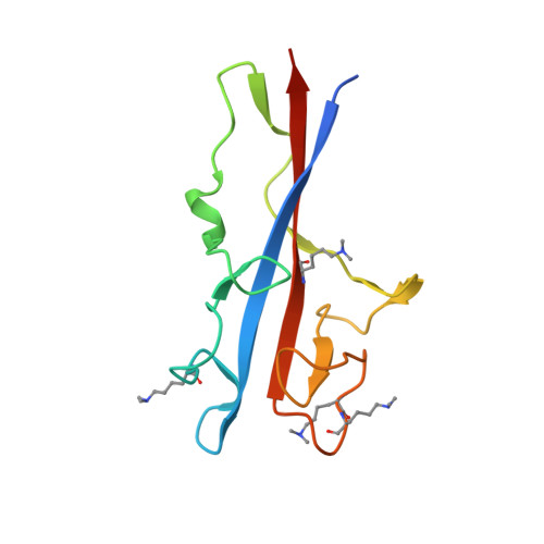
8P5P - PubMed Abstract:
Invasive bacteria enter the cytosol of host cells through initial uptake into bacteria-containing vacuoles (BCVs) and subsequent rupture of the BCV membrane, thereby exposing to the cytosol intraluminal, otherwise shielded danger signals such as glycans and sphingomyelin. The detection of glycans by galectin-8 triggers anti-bacterial autophagy, but how cells sense and respond to cytosolically exposed sphingomyelin remains unknown. Here, we identify TECPR1 (tectonin beta-propeller repeat containing 1) as a receptor for cytosolically exposed sphingomyelin, which recruits ATG5 into an E3 ligase complex that mediates lipid conjugation of LC3 independently of ATG16L1. TECPR1 binds sphingomyelin through its N-terminal DysF domain (N'DysF), a feature not shared by other mammalian DysF domains. Solving the crystal structure of N'DysF, we identified key residues required for the interaction, including a solvent-exposed tryptophan (W154) essential for binding to sphingomyelin-positive membranes and the conjugation of LC3 to lipids. Specificity of the ATG5/ATG12-E3 ligase responsible for the conjugation of LC3 is therefore conferred by interchangeable receptor subunits, that is, the canonical ATG16L1 and the sphingomyelin-specific TECPR1, in an arrangement reminiscent of certain multi-subunit ubiquitin E3 ligases.
- Division of Protein and Nucleic Acid Chemistry, MRC Laboratory of Molecular Biology, Cambridge, UK.
Organizational Affiliation: