Multi-modal cryo-EM reveals trimers of protein A10 to form the palisade layer in poxvirus cores.
Datler, J., Hansen, J.M., Thader, A., Schlogl, A., Bauer, L.W., Hodirnau, V.V., Schur, F.K.M.(2024) Nat Struct Mol Biol 31: 1114-1123
- PubMed: 38316877 Search on PubMedSearch on PubMed Central
- DOI: https://doi.org/10.1038/s41594-023-01201-6
- Primary Citation Related Structures:
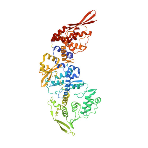
8P4K - PubMed Abstract:
Poxviruses are among the largest double-stranded DNA viruses, with members such as variola virus, monkeypox virus and the vaccination strain vaccinia virus (VACV). Knowledge about the structural proteins that form the viral core has remained sparse. While major core proteins have been annotated via indirect experimental evidence, their structures have remained elusive and they could not be assigned to individual core features. Hence, which proteins constitute which layers of the core, such as the palisade layer and the inner core wall, has remained enigmatic. Here we show, using a multi-modal cryo-electron microscopy (cryo-EM) approach in combination with AlphaFold molecular modeling, that trimers formed by the cleavage product of VACV protein A10 are the key component of the palisade layer. This allows us to place previously obtained descriptions of protein interactions within the core wall into perspective and to provide a detailed model of poxvirus core architecture. Importantly, we show that interactions within A10 trimers are likely generalizable over members of orthopox- and parapoxviruses.
- Institute of Science and Technology Austria (ISTA), Klosterneuburg, Austria.
Organizational Affiliation: