Fluorinated cGAMP analogs, which act as STING agonists and are not cleavable by poxins: Structural basis of their function.
Klima, M., Dejmek, M., Duchoslav, V., Eisenreichova, A., Sala, M., Chalupsky, K., Chalupska, D., Novotna, B., Birkus, G., Nencka, R., Boura, E.(2024) Structure 32: 433
- PubMed: 38325369 Search on PubMed
- DOI: https://doi.org/10.1016/j.str.2024.01.008
- Primary Citation Related Structures:
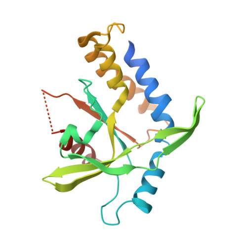
8ORV, 8ORW, 8P44, 8P45 - PubMed Abstract:
The cGAS-STING pathway is a crucial part of innate immunity; it serves to detect DNA in the cytoplasm and to defend against certain cancers, viruses, and bacteria. We designed and synthesized fluorinated carbocyclic cGAMP analogs, MD1203 and MD1202D (MDs), to enhance their stability and their affinity for STING. These compounds demonstrated exceptional activity against STING. Despite their distinct chemical modifications relative to the canonical cyclic dinucleotides (CDNs), crystallographic analysis revealed a binding mode with STING that was consistent with the canonical CDNs. Importantly, MDs were resistant to cleavage by viral poxin nucleases and MDs-bound poxin adopted an unliganded-like conformation. Moreover, MDs complexed with poxin showed a conformation distinct from cGAMP bound to poxin, closely resembling their conformation when bound to STING. In conclusion, the development of MD1203 and MD1202D showcases their potential as potent STING activators with remarkable stability against poxin-mediated degradation-a crucial characteristic for future development of antivirals.
- Institute of Organic Chemistry and Biochemistry AS CR, v.v.i., Flemingovo nam. 2., 166 10 Prague 6, Czech Republic.
Organizational Affiliation: