8P2N | pdb_00008p2n

Polymeric form of the BTB domain of ZBTB8A from Xenopus laevis


Experimental Data Snapshot

  • Method: X-RAY DIFFRACTION
  • Resolution: 3.10 Å
  • R-Value Free: 
    0.264 (Depositor), 0.266 (DCC) 
  • R-Value Work: 
    0.238 (Depositor), 0.243 (DCC) 
  • R-Value Observed: 
    0.240 (Depositor) 

Starting Model: in silico
View more details

wwPDB Validation 3D Report Full Report

Validation slider image for 8P2N

This is version 1.1 of the entry. See complete history

Literature

Dynamic BTB-domain filaments promote clustering of ZBTB proteins.

Mance, L.Bigot, N.Zhamungui Sanchez, E.Coste, F.Martin-Gonzalez, N.Zentout, S.Biliskov, M.Pukalo, Z.Mishra, A.Chapuis, C.Arteni, A.A.Lateur, A.Goffinont, S.Gaudon, V.Talhaoui, I.Casuso, I.Beaufour, M.Garnier, N.Artzner, F.Cadene, M.Huet, S.Castaing, B.Suskiewicz, M.J.

(2024) Mol Cell 84: 2490-2510.e9

  • DOI: https://doi.org/10.1016/j.molcel.2024.05.029
  • Primary Citation Related Structures: 
    8P2N, 8P2O, 8P2P, 8RIR, 8RIT

  • PubMed Abstract: 

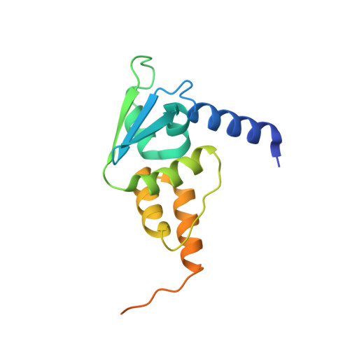
    The formation of dynamic protein filaments contributes to various biological functions by clustering individual molecules together and enhancing their binding to ligands. We report such a propensity for the BTB domains of certain proteins from the ZBTB family, a large eukaryotic transcription factor family implicated in differentiation and cancer. Working with Xenopus laevis and human proteins, we solved the crystal structures of filaments formed by dimers of the BTB domains of ZBTB8A and ZBTB18 and demonstrated concentration-dependent higher-order assemblies of these dimers in solution. In cells, the BTB-domain filamentation supports clustering of full-length human ZBTB8A and ZBTB18 into dynamic nuclear foci and contributes to the ZBTB18-mediated repression of a reporter gene. The BTB domains of up to 21 human ZBTB family members and two related proteins, NACC1 and NACC2, are predicted to behave in a similar manner. Our results suggest that filamentation is a more common feature of transcription factors than is currently appreciated.


  • Organizational Affiliation
    • Centre de Biophysique Moléculaire (CBM), UPR 4301, CNRS, affiliated with Université d'Orléans, 45071 Orléans Cedex 2, France.

Macromolecule Content 

  • Total Structure Weight: 35.18 kDa 
  • Atom Count: 1,815 
  • Modeled Residue Count: 246 
  • Deposited Residue Count: 312 
  • Unique protein chains: 1

Macromolecules

Find similar proteins by:|  3D Structure
Entity ID: 1
MoleculeChains  Sequence LengthOrganismDetailsImage
Zinc finger and BTB domain-containing protein 8A.1-A
A, B
156Xenopus laevisMutation(s): 0 
Gene Names: zbtb8a.1-azbtb8zbtb8.1
UniProt
Find proteins for Q0IH98 (Xenopus laevis)
Explore Q0IH98 
Go to UniProtKB:  Q0IH98
Entity Groups
Sequence Clusters30% Identity50% Identity70% Identity90% Identity95% Identity100% Identity
UniProt GroupQ0IH98
Sequence Annotations
Expand
Reference Sequence

Experimental Data & Validation

Experimental Data

  • Method: X-RAY DIFFRACTION
  • Resolution: 3.10 Å
  • R-Value Free:  0.264 (Depositor), 0.266 (DCC) 
  • R-Value Work:  0.238 (Depositor), 0.243 (DCC) 
  • R-Value Observed: 0.240 (Depositor) 
Space Group: P 41 3 2
Unit Cell:
Length ( Å )Angle ( ˚ )
a = 153.695α = 90
b = 153.695β = 90
c = 153.695γ = 90
Software Package:
Software NamePurpose
MxCuBEdata collection
XDSdata reduction
Aimlessdata scaling
PHASERphasing
Cootmodel building
PHENIXrefinement

Structure Validation

View Full Validation Report


Entry History 

& Funding Information

Deposition Data


Funding OrganizationLocationGrant Number
Centre National de la Recherche Scientifique (CNRS)France--

Revision History  (Full details and data files)

  • Version 1.0: 2024-06-05
    Type: Initial release
  • Version 1.1: 2024-12-18
    Changes: Database references, Refinement description, Structure summary