Characterization of two bacterial multi-flavinylated proteins harboring multiple covalent flavin cofactors.
Tong, Y., Rozeboom, H.J., Loonstra, M.R., Wijma, H.J., Fraaije, M.W.(2023) BBA Adv 4: 100097-100097
- PubMed: 37455753 Search on PubMedSearch on PubMed Central
- DOI: https://doi.org/10.1016/j.bbadva.2023.100097
- Primary Citation Related Structures:
8P2A, 8P2B - PubMed Abstract:
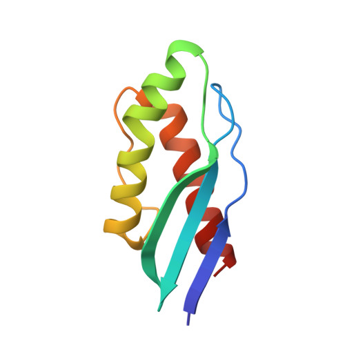
In recent years, studies have shown that a large number of bacteria secrete multi-flavinylated proteins. The exact roles and properties, of these extracellular flavoproteins that contain multiple covalently anchored FMN cofactors, are still largely unknown. Herein, we describe the biochemical and structural characterization of two multi-FMN-containing covalent flavoproteins, SaFMN3 from Streptomyces azureus and CbFMN4 from Clostridiaceae bacterium . Based on their primary structure, these proteins were predicted to contain three and four covalently tethered FMN cofactors, respectively. The genes encoding SaFMN3 and CbFMN4 were heterologously coexpressed with a flavin transferase (ApbE) in Escherichia coli , and could be purified by affinity chromatography in good yields. Both proteins were found to be soluble and to contain covalently bound FMN molecules. The SaFMN3 protein was studied in more detail and found to display a single redox potential (-184 mV) while harboring three covalently attached flavins. This is in line with the high sequence similarity when the domains of each flavoprotein are compared. The fully reduced form of SaFMN3 is able to use dioxygen as electron acceptor. Single domains from both proteins were expressed, purified and crystallized. The crystal structures were elucidated, which confirmed that the flavin cofactor is covalently attached to a threonine. Comparison of both crystal structures revealed a high similarity, even in the flavin binding pocket. Based on the crystal structure, mutants of the SaFMN3-D2 domain were designed to improve its fluorescence quantum yield by changing the microenvironment of the isoalloxazine moiety of the flavin cofactor. Residues that quench the flavin fluorescence were successfully identified. Our study reveals biochemical details of multi-FMN-containing proteins, contributing to a better understanding of their role in bacteria and providing leads to future utilization of these flavoprotein in biotechnology.
- Molecular Enzymology group, University of Groningen, Nijenborgh 4, Groningen 9747AG, the Netherlands.
Organizational Affiliation: