8P0P

Crystal structure of AaNGT complexed to UDP-2F-Glucose


Experimental Data Snapshot

  • Method: X-RAY DIFFRACTION
  • Resolution: 2.73 Å
  • R-Value Free: 0.245 
  • R-Value Work: 0.197 
  • R-Value Observed: 0.199 

wwPDB Validation   3D Report Full Report


Ligand Structure Quality Assessment 


This is version 1.1 of the entry. See complete history


Literature

Molecular basis for bacterial N-glycosylation by a soluble HMW1C-like N-glycosyltransferase.

Piniello, B.Macias-Leon, J.Miyazaki, S.Garcia-Garcia, A.Companon, I.Ghirardello, M.Taleb, V.Veloz, B.Corzana, F.Miyagawa, A.Rovira, C.Hurtado-Guerrero, R.

(2023) Nat Commun 14: 5785-5785

  • DOI: https://doi.org/10.1038/s41467-023-41238-1
  • Primary Citation of Related Structures:  
    8P0O, 8P0P, 8P0Q

  • PubMed Abstract: 

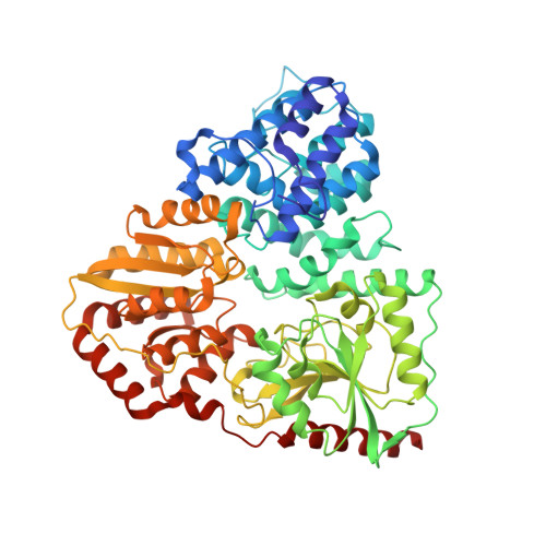
    Soluble HMW1C-like N-glycosyltransferases (NGTs) catalyze the glycosylation of Asn residues in proteins, a process fundamental for bacterial autoaggregation, adhesion and pathogenicity. However, our understanding of their molecular mechanisms is hindered by the lack of structures of enzymatic complexes. Here, we report structures of binary and ternary NGT complexes of Aggregatibacter aphrophilus NGT (AaNGT), revealing an essential dyad of basic/acidic residues located in the N-terminal all α-domain (AAD) that intimately recognizes the Thr residue within the conserved motif Asn 0 -X +1 -Ser/Thr +2 . Poor substrates and inhibitors such as UDP-galactose and UDP-glucose mimetics adopt non-productive conformations, decreasing or impeding catalysis. QM/MM simulations rationalize these results, showing that AaNGT follows a S N 2 reaction mechanism in which the acceptor asparagine uses its imidic form for catalysis and the UDP-glucose phosphate group acts as a general base. These findings provide key insights into the mechanism of NGTs and will facilitate the design of structure-based inhibitors to treat diseases caused by non-typeable H. influenzae or other Gram-negative bacteria.


  • Organizational Affiliation

    Departament de Química Inorgànica i Orgànica (Secció de Química Orgànica) and Institut de Química Teòrica i Computacional (IQTCUB), Universitat de Barcelona, Martí i Franquès 1, 08028, Barcelona, Spain.


Macromolecules
Find similar proteins by:  (by identity cutoff)  |  3D Structure
Entity ID: 1
MoleculeChains Sequence LengthOrganismDetailsImage
Adhesin
A, B
622Aggregatibacter aphrophilusMutation(s): 0 
Gene Names: DOL88_00785
UniProt
Find proteins for A0A3M6PNT1 (Aggregatibacter aphrophilus)
Explore A0A3M6PNT1 
Go to UniProtKB:  A0A3M6PNT1
Entity Groups  
Sequence Clusters30% Identity50% Identity70% Identity90% Identity95% Identity100% Identity
UniProt GroupA0A3M6PNT1
Sequence Annotations
Expand
  • Reference Sequence
Small Molecules
Ligands 2 Unique
IDChains Name / Formula / InChI Key2D Diagram3D Interactions
U2F (Subject of Investigation/LOI)
Query on U2F

Download Ideal Coordinates CCD File 
C [auth A]URIDINE-5'-DIPHOSPHATE-2-DEOXY-2-FLUORO-ALPHA-D-GLUCOSE
C15 H23 F N2 O16 P2
NGTCPFGWXMBZEP-NQQHDEILSA-N
UDP (Subject of Investigation/LOI)
Query on UDP

Download Ideal Coordinates CCD File 
D [auth B]URIDINE-5'-DIPHOSPHATE
C9 H14 N2 O12 P2
XCCTYIAWTASOJW-XVFCMESISA-N
Experimental Data & Validation

Experimental Data

  • Method: X-RAY DIFFRACTION
  • Resolution: 2.73 Å
  • R-Value Free: 0.245 
  • R-Value Work: 0.197 
  • R-Value Observed: 0.199 
  • Space Group: P 21 21 21
Unit Cell:
Length ( Å )Angle ( ˚ )
a = 47.068α = 90
b = 114.005β = 90
c = 258.008γ = 90
Software Package:
Software NamePurpose
REFMACrefinement
XDSdata reduction
SCALAdata scaling
PHASERphasing

Structure Validation

View Full Validation Report


Ligand Structure Quality Assessment 


Entry History & Funding Information

Deposition Data


Funding OrganizationLocationGrant Number
Ministry of Economy and Competitiveness (MINECO)SpainPID2019-105451GB-I00

Revision History  (Full details and data files)

  • Version 1.0: 2023-09-06
    Type: Initial release
  • Version 1.1: 2023-09-27
    Changes: Database references