Integrated NMR-crystallography-computational approach for molecular recognition studies of human G alpha i3 protein by a small molecule inhibitor.
Ferreras-Gutierrez, M., Minguez-Toral, M., Ibanez de Opakua, A., Martin-Santamaria, S., Garcia-Marcos, M., Medrano, F.J., Blanco, F.J.(2024) Int J Biol Macromol 290: 138977-138977
- PubMed: 39706421 Search on PubMed
- DOI: https://doi.org/10.1016/j.ijbiomac.2024.138977
- Primary Citation Related Structures:
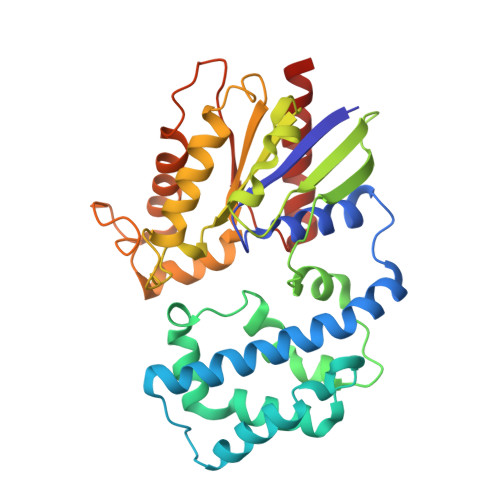
8OY1 - PubMed Abstract:
The small molecule IGGi-11 targets Gαi subunits of heterotrimeric guanine nucleotide-binding proteins. Gα subunits are activated by G-protein-coupled receptors in response to extracellular stimuli by accelerating the exchange of GDP for GTP, but they are also activated by intracellular proteins like GIV, of which elevated levels correlate with increased cell migration and cancer metastasis. IGGi-11 disrupts the interaction of Gαi proteins with GIV and inhibits pro-invasive traits of metastatic breast cancer cells without interfering with GPCR signaling. IGGi-11 is a competitive inhibitor but binds Gαi3 with a 10-fold lower affinity than GIV. To guide the design of higher affinity inhibitors, we aimed at obtaining high-resolution structural data on the complex. To facilitate its crystallization, we have removed the most flexible residues at the chain ends of Gαi3, identified by NMR. While Gαi3 crystals grown with excess IGGi-11 did not show the bound compound, computational docking and molecular dynamics simulations identified the interactions driving the molecular recognition. This approach revealed heterogeneous binding due to the symmetry of IGGi-11 chemical structure and to the elongated shape and flexibility of the binding site. Our results suggest that chemical modifications breaking IGGi-11 symmetry might yield inhibitors with higher affinity and potential as antimetastatic drugs.
- Centro de Investigaciones Biológicas Margarita Salas (CIB), CSIC, Madrid 28040, Spain.
Organizational Affiliation: