8OX6 | pdb_00008ox6

Cryo-EM structure of ATP8B1-CDC50A in E1P conformation


Experimental Data Snapshot

  • Method: ELECTRON MICROSCOPY
  • Resolution: 2.39 Å
  • Aggregation State: PARTICLE 
  • Reconstruction Method: SINGLE PARTICLE 

Starting Model: experimental
View more details

wwPDB Validation 3D Report Full Report

Validation slider image for 8OX6

This is version 1.1 of the entry. See complete history

Literature

Activation and substrate specificity of the human P4-ATPase ATP8B1.

Dieudonne, T.Kummerer, F.Laursen, M.J.Stock, C.Flygaard, R.K.Khalid, S.Lenoir, G.Lyons, J.A.Lindorff-Larsen, K.Nissen, P.

(2023) Nat Commun 14: 7492-7492

  • DOI: https://doi.org/10.1038/s41467-023-42828-9
  • Primary Citation Related Structures: 
    8OX4, 8OX5, 8OX6, 8OX7, 8OX8, 8OX9, 8OXA, 8OXB, 8OXC

  • PubMed Abstract: 

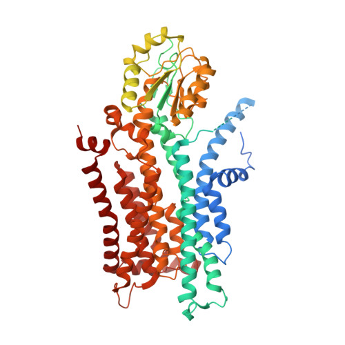
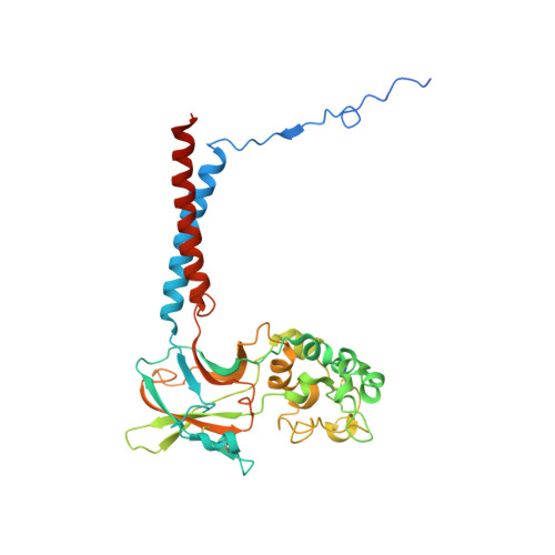
    Asymmetric distribution of phospholipids in eukaryotic membranes is essential for cell integrity, signaling pathways, and vesicular trafficking. P4-ATPases, also known as flippases, participate in creating and maintaining this asymmetry through active transport of phospholipids from the exoplasmic to the cytosolic leaflet. Here, we present a total of nine cryo-electron microscopy structures of the human flippase ATP8B1-CDC50A complex at 2.4 to 3.1 Å overall resolution, along with functional and computational studies, addressing the autophosphorylation steps from ATP, substrate recognition and occlusion, as well as a phosphoinositide binding site. We find that the P4-ATPase transport site is occupied by water upon phosphorylation from ATP. Additionally, we identify two different autoinhibited states, a closed and an outward-open conformation. Furthermore, we identify and characterize the PI(3,4,5)P 3 binding site of ATP8B1 in an electropositive pocket between transmembrane segments 5, 7, 8, and 10. Our study also highlights the structural basis of a broad lipid specificity of ATP8B1 and adds phosphatidylinositol as a transport substrate for ATP8B1. We report a critical role of the sn-2 ester bond of glycerophospholipids in substrate recognition by ATP8B1 through conserved S403. These findings provide fundamental insights into ATP8B1 catalytic cycle and regulation, and substrate recognition in P4-ATPases.


  • Organizational Affiliation
    • DANDRITE, Nordic EMBL Partnership for Molecular Medicine, Department of Molecular Biology and Genetics, Aarhus University, Aarhus, Denmark. thibaud.dieudonne@i2bc.paris-saclay.fr.

Macromolecule Content 

  • Total Structure Weight: 178.53 kDa 
  • Atom Count: 7,359 
  • Modeled Residue Count: 903 
  • Deposited Residue Count: 1,550 
  • Unique protein chains: 2

Macromolecules

Find similar proteins by:|  3D Structure
Entity ID: 1
MoleculeChains  Sequence LengthOrganismDetailsImage
Phospholipid-transporting ATPase IC1,185Homo sapiensMutation(s): 1 
Gene Names: ATP8B1ATPICFIC1PFIC
EC: 7.6.2.1
Membrane Entity: Yes 
UniProt & NIH Common Fund Data Resources
Find proteins for O43520 (Homo sapiens)
Explore O43520 
Go to UniProtKB:  O43520
PHAROS:  O43520
GTEx:  ENSG00000081923 
Entity Groups
Sequence Clusters30% Identity50% Identity70% Identity90% Identity95% Identity100% Identity
UniProt GroupO43520
Sequence Annotations
Expand
Reference Sequence
Find similar proteins by:|  3D Structure
Entity ID: 2
MoleculeChains  Sequence LengthOrganismDetailsImage
Cell cycle control protein 50A365Homo sapiensMutation(s): 0 
Gene Names: TMEM30AC6orf67CDC50A
Membrane Entity: Yes 
UniProt & NIH Common Fund Data Resources
Find proteins for Q9NV96 (Homo sapiens)
Explore Q9NV96 
Go to UniProtKB:  Q9NV96
PHAROS:  Q9NV96
GTEx:  ENSG00000112697 
Entity Groups
Sequence Clusters30% Identity50% Identity70% Identity90% Identity95% Identity100% Identity
UniProt GroupQ9NV96
Glycosylation
Glycosylation Sites: 3Go to GlyGen: Q9NV96-1
Sequence Annotations
Expand
Reference Sequence

Oligosaccharides

Help  
Entity ID: 3
MoleculeChains Length2D Diagram GlycosylationD Interactions
beta-D-mannopyranose-(1-4)-2-acetamido-2-deoxy-beta-D-glucopyranose-(1-4)-2-acetamido-2-deoxy-beta-D-glucopyranose
C
3N-Glycosylation
Glycosylation Resources
GlyTouCan: G15407YE
GlyCosmos: G15407YE
GlyGen: G15407YE

Experimental Data & Validation

Experimental Data

  • Method: ELECTRON MICROSCOPY
  • Resolution: 2.39 Å
  • Aggregation State: PARTICLE 
  • Reconstruction Method: SINGLE PARTICLE 
EM Software:
TaskSoftware PackageVersion
MODEL REFINEMENTPHENIX
RECONSTRUCTIONcryoSPARC3

Structure Validation

View Full Validation Report


Entry History 

& Funding Information

Deposition Data


Funding OrganizationLocationGrant Number
H2020 Marie Curie Actions of the European CommissionEuropean Union101024542
LundbeckfondenDenmarkR310-2018-3713
LundbeckfondenDenmark155-2015-2666
LundbeckfondenDenmarkR335-2019-2053
Agence Nationale de la Recherche (ANR)FranceANR-14-CE09-0022
French Infrastructure for Integrated Structural Biology (FRISBI)FranceANR-10-INSB-05
Engineering and Physical Sciences Research CouncilUnited KingdomEP/X035603
Engineering and Physical Sciences Research CouncilUnited KingdomEP/C030779

Revision History  (Full details and data files)

  • Version 1.0: 2023-11-29
    Type: Initial release
  • Version 1.1: 2024-11-13
    Changes: Data collection, Structure summary