8OUO | pdb_00008ouo

Human TPC2 in Complex with Antagonist (S)-SG-094


Experimental Data Snapshot

  • Method: ELECTRON MICROSCOPY
  • Resolution: 3.00 Å
  • Aggregation State: PARTICLE 
  • Reconstruction Method: SINGLE PARTICLE 

wwPDB Validation 3D Report Full Report

Validation slider image for 8OUO

This is version 1.3 of the entry. See complete history

Literature

Structural basis for inhibition of the lysosomal two-pore channel TPC2 by a small molecule antagonist.

Chi, G.Jaslan, D.Kudrina, V.Bock, J.Li, H.Pike, A.C.W.Rautenberg, S.Krogsaeter, E.Bohstedt, T.Wang, D.McKinley, G.Fernandez-Cid, A.Mukhopadhyay, S.M.M.Burgess-Brown, N.A.Keller, M.Bracher, F.Grimm, C.Durr, K.L.

(2024) Structure 32: 1137-1149.e4

  • DOI: https://doi.org/10.1016/j.str.2024.05.005
  • Primary Citation Related Structures: 
    8OUO

  • PubMed Abstract: 

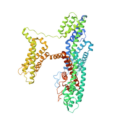
    Two pore channels are lysosomal cation channels with crucial roles in tumor angiogenesis and viral release from endosomes. Inhibition of the two-pore channel 2 (TPC2) has emerged as potential therapeutic strategy for the treatment of cancers and viral infections, including Ebola and COVID-19. Here, we demonstrate that antagonist SG-094, a synthetic analog of the Chinese alkaloid medicine tetrandrine with increased potency and reduced toxicity, induces asymmetrical structural changes leading to a single binding pocket at only one intersubunit interface within the asymmetrical dimer. Supported by functional characterization of mutants by Ca 2+ imaging and patch clamp experiments, we identify key residues in S1 and S4 involved in compound binding to the voltage sensing domain II. SG-094 arrests IIS4 in a downward shifted state which prevents pore opening via the IIS4/S5 linker, hence resembling gating modifiers of canonical VGICs. These findings may guide the rational development of new therapeutics antagonizing TPC2 activity.


  • Organizational Affiliation
    • Centre for Medicines Discovery, Nuffield Department of Medicine, University of Oxford, Nuffield Department of Medicine Research Building, Oxford OX3 7FZ, UK; Structural Genomics Consortium, Nuffield Department of Medicine, University of Oxford, Nuffield Department of Medicine Research Building, Oxford OX3 7FZ, UK. Electronic address: gamma.chi@cmd.ox.ac.uk.

Macromolecule Content 

  • Total Structure Weight: 180.66 kDa 
  • Atom Count: 10,749 
  • Modeled Residue Count: 1,273 
  • Deposited Residue Count: 1,504 
  • Unique protein chains: 1

Macromolecules

Find similar proteins by:|  3D Structure
Entity ID: 1
MoleculeChains  Sequence LengthOrganismDetailsImage
Two pore channel protein 2
A, B
752Homo sapiensMutation(s): 2 
Gene Names: TPCN2TPC2
Membrane Entity: Yes 
UniProt & NIH Common Fund Data Resources
Find proteins for Q8NHX9 (Homo sapiens)
Explore Q8NHX9 
Go to UniProtKB:  Q8NHX9
PHAROS:  Q8NHX9
GTEx:  ENSG00000162341 
Entity Groups
Sequence Clusters30% Identity50% Identity70% Identity90% Identity95% Identity100% Identity
UniProt GroupQ8NHX9
Sequence Annotations
Expand
Reference Sequence

Small Molecules

Ligands 5 Unique
IDChains Name / Formula / InChI Key2D Diagram3D Interactions
Q7G

Query on Q7G


Download:Ideal Coordinates CCD File
D [auth A],
J [auth A],
P [auth B]
2-{[(4-O-alpha-D-glucopyranosyl-alpha-D-glucopyranosyl)oxy]methyl}-4-{[(3beta,9beta,14beta,17beta,25R)-spirost-5-en-3-yl]oxy}butyl 4-O-alpha-D-glucopyranosyl-alpha-D-glucopyranoside
C56 H92 O25
LKBFXDNKNZXHHW-NXLTVWPKSA-N
PLD

Query on PLD


Download:Ideal Coordinates CCD File
C [auth A],
K [auth B]
di-heneicosanoyl phosphatidyl choline
C50 H101 N O8 P
QFFSGJSMHPWZOB-QSCHNALKSA-O
PCF

Query on PCF


Download:Ideal Coordinates CCD File
F [auth A],
L [auth B]
1,2-DIACYL-SN-GLYCERO-3-PHOSHOCHOLINE
C40 H80 N O8 P
KILNVBDSWZSGLL-KXQOOQHDSA-N
Y01

Query on Y01


Download:Ideal Coordinates CCD File
G [auth A]
H [auth A]
I [auth A]
M [auth B]
N [auth B]
G [auth A],
H [auth A],
I [auth A],
M [auth B],
N [auth B],
O [auth B]
CHOLESTEROL HEMISUCCINATE
C31 H50 O4
WLNARFZDISHUGS-MIXBDBMTSA-N
W4E
(Subject of Investigation/LOI)

Query on W4E


Download:Ideal Coordinates CCD File
E [auth A](1S)-6-methoxy-2-methyl-7-phenoxy-1-[(4-phenoxyphenyl)methyl]-3,4-dihydro-1H-isoquinoline
C30 H29 N O3
BTNHPUSFPKFAPU-NDEPHWFRSA-N

Experimental Data & Validation

Experimental Data

  • Method: ELECTRON MICROSCOPY
  • Resolution: 3.00 Å
  • Aggregation State: PARTICLE 
  • Reconstruction Method: SINGLE PARTICLE 
EM Software:
TaskSoftware PackageVersion
MODEL REFINEMENTPHENIX

Structure Validation

View Full Validation Report


Entry History 

& Funding Information

Deposition Data


Funding OrganizationLocationGrant Number
Wellcome TrustUnited Kingdom--

Revision History  (Full details and data files)

  • Version 1.0: 2024-06-12
    Type: Initial release
  • Version 1.1: 2024-08-21
    Changes: Data collection, Database references
  • Version 1.2: 2024-10-23
    Changes: Data collection, Structure summary
  • Version 1.3: 2025-07-02
    Changes: Data collection