8OSP

GCN5-related N-Acetyltransferase from Lactobacillus curiae


Experimental Data Snapshot

  • Method: X-RAY DIFFRACTION
  • Resolution: 1.95 Å
  • R-Value Free: 0.221 
  • R-Value Work: 0.183 
  • R-Value Observed: 0.185 

wwPDB Validation   3D Report Full Report


This is version 1.1 of the entry. See complete history


Literature

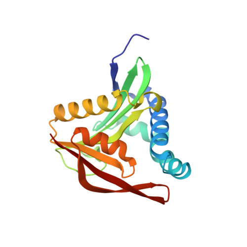
Crystal structure of a GCN5-related N-acetyltransferase from Lactobacillus curiae.

Fleming, J.R.Hauth, F.Hartig, J.S.Mayans, O.

(2023) Acta Crystallogr F Struct Biol Commun 79: 217-223

  • DOI: https://doi.org/10.1107/S2053230X2300571X
  • Primary Citation of Related Structures:  
    8OSP

  • PubMed Abstract: 

    Members of the GCN5-related N-acetyltransferase (GNAT) family are found in all domains of life and are involved in processes ranging from protein synthesis and gene expression to detoxification and virulence. Due to the variety of their macromolecular targets, GNATs are a highly diverse family of proteins. Currently, 3D structures of only a small number of GNAT representatives are available and thus the family remains poorly characterized. Here, the crystal structure of the guanidine riboswitch-associated GNAT from Lactobacillus curiae (LcGNAT) that acetylates canavanine, a structural analogue of arginine with antimetabolite properties, is reported. LcGNAT shares the conserved fold of the members of the GNAT superfamily, but does not contain an N-terminal β0 strand and instead contains a C-terminal β7 strand. Its P-loop, which coordinates the pyrophosphate moiety of the acetyl-coenzyme A cosubstrate, is degenerated. These features are shared with its closest homologues in the polyamine acetyltransferase subclass. Site-directed mutagenesis revealed a central role of the conserved residue Tyr142 in catalysis, as well as the semi-conserved Tyr97 and Glu92, suggesting that despite its individual substrate specificity LcGNAT performs the classical reaction mechanism of this family.


  • Organizational Affiliation

    Department of Biology, University of Konstanz, Universitätsstrasse 10, 78457 Konstanz, Germany.


Macromolecules
Find similar proteins by:  (by identity cutoff)  |  3D Structure
Entity ID: 1
MoleculeChains Sequence LengthOrganismDetailsImage
N-acetyltransferaseA,
B [auth C]
174LactobacillusMutation(s): 0 
Gene Names: PL11_000275
UniProt
Find proteins for A0A1S6QFV5 (Lentilactobacillus curieae)
Explore A0A1S6QFV5 
Go to UniProtKB:  A0A1S6QFV5
Entity Groups  
Sequence Clusters30% Identity50% Identity70% Identity90% Identity95% Identity100% Identity
UniProt GroupA0A1S6QFV5
Sequence Annotations
Expand
  • Reference Sequence
Small Molecules
Ligands 3 Unique
IDChains Name / Formula / InChI Key2D Diagram3D Interactions
MES
Query on MES

Download Ideal Coordinates CCD File 
D [auth A],
E [auth A],
K [auth C],
L [auth C]
2-(N-MORPHOLINO)-ETHANESULFONIC ACID
C6 H13 N O4 S
SXGZJKUKBWWHRA-UHFFFAOYSA-N
PO4
Query on PO4

Download Ideal Coordinates CCD File 
F [auth A],
M [auth C]
PHOSPHATE ION
O4 P
NBIIXXVUZAFLBC-UHFFFAOYSA-K
GOL
Query on GOL

Download Ideal Coordinates CCD File 
C [auth A],
G [auth A],
H [auth A],
I [auth A],
J [auth C]
GLYCEROL
C3 H8 O3
PEDCQBHIVMGVHV-UHFFFAOYSA-N
Experimental Data & Validation

Experimental Data

  • Method: X-RAY DIFFRACTION
  • Resolution: 1.95 Å
  • R-Value Free: 0.221 
  • R-Value Work: 0.183 
  • R-Value Observed: 0.185 
  • Space Group: P 1 21 1
Unit Cell:
Length ( Å )Angle ( ˚ )
a = 60.68α = 90
b = 37.08β = 97.67
c = 83.65γ = 90
Software Package:
Software NamePurpose
PHENIXrefinement
XDSdata reduction
XSCALEdata scaling
PHENIXphasing

Structure Validation

View Full Validation Report


Entry History & Funding Information

Deposition Data


Funding OrganizationLocationGrant Number
Not funded--

Revision History  (Full details and data files)

  • Version 1.0: 2023-07-05
    Type: Initial release
  • Version 1.1: 2023-08-23
    Changes: Data collection, Database references