8ORP | pdb_00008orp

Crystal structure of Drosophila melanogaster alpha-amylase in complex with the inhibitor acarbose


Experimental Data Snapshot

  • Method: X-RAY DIFFRACTION
  • Resolution: 2.00 Å
  • R-Value Free: 
    0.212 (Depositor), 0.212 (DCC) 
  • R-Value Work: 
    0.174 (Depositor), 0.174 (DCC) 
  • R-Value Observed: 
    0.176 (Depositor) 

Starting Model: experimental
View more details

wwPDB Validation 3D Report Full Report

Validation slider image for 8ORP

This is version 1.1 of the entry. See complete history

Literature

Structural and Functional Characterization of Drosophila melanogaster alpha-Amylase.

Rhimi, M.Da Lage, J.L.Haser, R.Feller, G.Aghajari, N.

(2023) Molecules 28

  • DOI: https://doi.org/10.3390/molecules28145327
  • Primary Citation Related Structures: 
    8OR6, 8ORP

  • PubMed Abstract: 

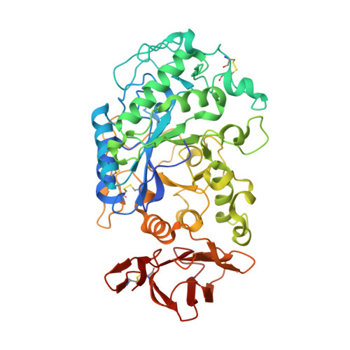
    Insects rely on carbohydrates such as starch and glycogen as an energy supply for growth of larvae and for longevity. In this sense α-amylases have essential roles under extreme conditions, e.g., during nutritional or temperature stress, thereby contributing to survival of the insect. This makes them interesting targets for combating insect pests. Drosophila melanogaster α-amylase, DMA, which belongs to the glycoside hydrolase family 13, sub family 15, has been studied from an evolutionary, biochemical, and structural point of view. Our studies revealed that the DMA enzyme is active over a broad temperature and pH range, which is in agreement with the fluctuating environmental changes with which the insect is confronted. Crystal structures disclosed a new nearly fully solvated metal ion, only coordinated to the protein via Gln263. This residue is only conserved in the subgroup of D. melanogaster and may thus contribute to the enzyme adaptive response to large temperature variations. Studies of the effect of plant inhibitors and the pseudo-tetrasaccharide inhibitor acarbose on DMA activity, allowed us to underline the important role of the so-called flexible loop on activity/inhibition, but also to suggest that the inhibition modes of the wheat inhibitors WI-1 and WI-3 on DMA, are likely different.


  • Organizational Affiliation
    • Molecular Microbiology and Structural Biochemistry, UMR5086, CNRS, University of Lyon 1, 7 Passage du Vercors, F-69367 Lyon, CEDEX 07, France.

Macromolecule Content 

  • Total Structure Weight: 112.64 kDa 
  • Atom Count: 8,503 
  • Modeled Residue Count: 952 
  • Deposited Residue Count: 988 
  • Unique protein chains: 1

Macromolecules

Find similar proteins by:|  3D Structure
Entity ID: 1
MoleculeChains  Sequence LengthOrganismDetailsImage
Alpha-amylase A
A, B
494Drosophila melanogasterMutation(s): 0 
Gene Names: Amy-pAmyACG18730
EC: 3.2.1.1
UniProt
Find proteins for P08144 (Drosophila melanogaster)
Explore P08144 
Go to UniProtKB:  P08144
Entity Groups
Sequence Clusters30% Identity50% Identity70% Identity90% Identity95% Identity100% Identity
UniProt GroupP08144
Sequence Annotations
Expand
Reference Sequence

Oligosaccharides

Help  
Entity ID: 2
MoleculeChains Length2D Diagram GlycosylationD Interactions
4,6-dideoxy-4-{[(1S,2S,3S,4R,5R)-2,3,4-trihydroxy-5-(hydroxymethyl)cyclohexyl]amino}-alpha-D-glucopyranose-(1-4)-alpha-D-glucopyranose-(1-4)-beta-D-glucopyranose
C, E
3N/AN/A
Glycosylation Resources
GlyTouCan: G45133CV
GlyCosmos: G45133CV
Entity ID: 3
MoleculeChains Length2D Diagram GlycosylationD Interactions
4,6-dideoxy-4-{[(1S,2S,3S,4R,5R)-2,3,4-trihydroxy-5-(hydroxymethyl)cyclohexyl]amino}-alpha-D-glucopyranose-(1-4)-alpha-D-glucopyranose
D, F
2N/AN/A

Small Molecules

Ligands 3 Unique
IDChains Name / Formula / InChI Key2D Diagram3D Interactions
SR
(Subject of Investigation/LOI)

Query on SR


Download:Ideal Coordinates CCD File
AA [auth A],
WA [auth B],
XA [auth B],
Z [auth A]
STRONTIUM ION
Sr
PWYYWQHXAPXYMF-UHFFFAOYSA-N
EDO

Query on EDO


Download:Ideal Coordinates CCD File
CA [auth B]
DA [auth B]
EA [auth B]
FA [auth B]
GA [auth B]
CA [auth B],
DA [auth B],
EA [auth B],
FA [auth B],
GA [auth B],
H [auth A],
HA [auth B],
I [auth A],
IA [auth B],
J [auth A],
JA [auth B],
K [auth A],
KA [auth B],
L [auth A],
LA [auth B],
M [auth A],
MA [auth B],
N [auth A],
NA [auth B],
O [auth A],
OA [auth B],
P [auth A],
PA [auth B],
Q [auth A],
QA [auth B],
R [auth A],
RA [auth B],
S [auth A],
SA [auth B],
T [auth A],
TA [auth B],
U [auth A],
UA [auth B],
V [auth A],
VA [auth B],
W [auth A],
X [auth A],
Y [auth A]
1,2-ETHANEDIOL
C2 H6 O2
LYCAIKOWRPUZTN-UHFFFAOYSA-N
CL
(Subject of Investigation/LOI)

Query on CL


Download:Ideal Coordinates CCD File
BA [auth B],
G [auth A]
CHLORIDE ION
Cl
VEXZGXHMUGYJMC-UHFFFAOYSA-M

Experimental Data & Validation

Experimental Data

  • Method: X-RAY DIFFRACTION
  • Resolution: 2.00 Å
  • R-Value Free:  0.212 (Depositor), 0.212 (DCC) 
  • R-Value Work:  0.174 (Depositor), 0.174 (DCC) 
  • R-Value Observed: 0.176 (Depositor) 
Space Group: P 1 21 1
Unit Cell:
Length ( Å )Angle ( ˚ )
a = 80.08α = 90
b = 72.54β = 98.41
c = 98.21γ = 90
Software Package:
Software NamePurpose
PHENIXrefinement
XDSdata reduction
XSCALEdata scaling
PHENIXphasing

Structure Validation

View Full Validation Report


Entry History 

& Funding Information

Deposition Data


Funding OrganizationLocationGrant Number
Centre National de la Recherche Scientifique (CNRS)France--

Revision History  (Full details and data files)

  • Version 1.0: 2023-08-16
    Type: Initial release
  • Version 1.1: 2025-10-01
    Changes: Advisory, Derived calculations, Structure summary