8ORO

CRYSTAL STRUCTURE OF THE COFACTOR-DEVOID 1-H-3-HYDROXY-4- OXOQUINALDINE 2,4-DIOXYGENASE (HOD) S101A VARIANT COMPLEXED WITH 2-METHYL-QUINOLIN-4(1H)-ONE UNDER HYPEROXYC CONDITIONS


Experimental Data Snapshot

  • Method: X-RAY DIFFRACTION
  • Resolution: 2.00 Å
  • R-Value Free: 0.269 
  • R-Value Work: 0.227 

wwPDB Validation   3D Report Full Report


Ligand Structure Quality Assessment 


This is version 1.0 of the entry. See complete history


Literature

Evolutionary adaptation from hydrolytic to oxygenolytic catalysis at the alpha / beta-hydrolase fold.

Bui, S.Gil-Guerrero, S.van der Linden, P.Carpentier, P.Ceccarelli, M.Jambrina, P.G.Steiner, R.A.

(2023) Chem Sci 14: 10547-10560

  • DOI: https://doi.org/10.1039/d3sc03044j
  • Primary Citation of Related Structures:  
    7OJM, 7OKZ, 8A97, 8ORO, 8OXN, 8OXT

  • PubMed Abstract: 

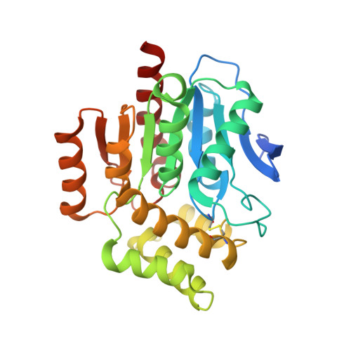
    Protein fold adaptation to novel enzymatic reactions is a fundamental evolutionary process. Cofactor-independent oxygenases degrading N -heteroaromatic substrates belong to the α/β-hydrolase (ABH) fold superfamily that typically does not catalyze oxygenation reactions. Here, we have integrated crystallographic analyses under normoxic and hyperoxic conditions with molecular dynamics and quantum mechanical calculations to investigate its prototypic 1- H -3-hydroxy-4-oxoquinaldine 2,4-dioxygenase (HOD) member. O 2 localization to the "oxyanion hole", where catalysis occurs, is an unfavorable event and the direct competition between dioxygen and water for this site is modulated by the "nucleophilic elbow" residue. A hydrophobic pocket that overlaps with the organic substrate binding site can act as a proximal dioxygen reservoir. Freeze-trap pressurization allowed the structure of the ternary complex with a substrate analogue and O 2 bound at the oxyanion hole to be determined. Theoretical calculations reveal that O 2 orientation is coupled to the charge of the bound organic ligand. When 1- H -3-hydroxy-4-oxoquinaldine is uncharged, O 2 binds with its molecular axis along the ligand's C2-C4 direction in full agreement with the crystal structure. Substrate activation triggered by deprotonation of its 3-OH group by the His-Asp dyad, rotates O 2 by approximately 60°. This geometry maximizes the charge transfer between the substrate and O 2 , thus weakening the double bond of the latter. Electron density transfer to the O 2 (π*) orbital promotes the formation of the peroxide intermediate via intersystem crossing that is rate-determining. Our work provides a detailed picture of how evolution has repurposed the ABH-fold architecture and its simple catalytic machinery to accomplish metal-independent oxygenation.


  • Organizational Affiliation

    Randall Centre for Cell and Molecular Biophysics, King's College London London SE1 1UL UK roberto.steiner@kcl.ac.uk.


Macromolecules
Find similar proteins by:  (by identity cutoff)  |  3D Structure
Entity ID: 1
MoleculeChains Sequence LengthOrganismDetailsImage
1H-3-hydroxy-4-oxoquinaldine 2,4-dioxygenaseA [auth AAA],
B [auth BBB]
288Paenarthrobacter nitroguajacolicusMutation(s): 2 
Gene Names: hodmeqEARUE_113p00080pAL1.008
EC: 1.13.11.48
UniProt
Find proteins for O31266 (Paenarthrobacter nitroguajacolicus)
Explore O31266 
Go to UniProtKB:  O31266
Entity Groups  
Sequence Clusters30% Identity50% Identity70% Identity90% Identity95% Identity100% Identity
UniProt GroupO31266
Sequence Annotations
Expand
  • Reference Sequence
Small Molecules
Ligands 5 Unique
IDChains Name / Formula / InChI Key2D Diagram3D Interactions
VFH (Subject of Investigation/LOI)
Query on VFH

Download Ideal Coordinates CCD File 
C [auth AAA],
G [auth BBB]
2-methyl-quinolin-4(1H)-one
C10 H9 N O
NWINIEGDLHHNLH-UHFFFAOYSA-N
TAR
Query on TAR

Download Ideal Coordinates CCD File 
E [auth AAA],
F [auth AAA],
I [auth BBB],
J [auth BBB]
D(-)-TARTARIC ACID
C4 H6 O6
FEWJPZIEWOKRBE-LWMBPPNESA-N
GOL
Query on GOL

Download Ideal Coordinates CCD File 
K [auth BBB]GLYCEROL
C3 H8 O3
PEDCQBHIVMGVHV-UHFFFAOYSA-N
OXY (Subject of Investigation/LOI)
Query on OXY

Download Ideal Coordinates CCD File 
D [auth AAA],
H [auth BBB]
OXYGEN MOLECULE
O2
MYMOFIZGZYHOMD-UHFFFAOYSA-N
NA
Query on NA

Download Ideal Coordinates CCD File 
L [auth BBB]SODIUM ION
Na
FKNQFGJONOIPTF-UHFFFAOYSA-N
Experimental Data & Validation

Experimental Data

  • Method: X-RAY DIFFRACTION
  • Resolution: 2.00 Å
  • R-Value Free: 0.269 
  • R-Value Work: 0.227 
  • Space Group: P 41
Unit Cell:
Length ( Å )Angle ( ˚ )
a = 120.11α = 90
b = 120.11β = 90
c = 44.82γ = 90
Software Package:
Software NamePurpose
REFMACrefinement
XDSdata reduction
XSCALEdata scaling
PHASERphasing

Structure Validation

View Full Validation Report


Ligand Structure Quality Assessment 


Entry History & Funding Information

Deposition Data


Funding OrganizationLocationGrant Number
Biotechnology and Biological Sciences Research Council (BBSRC)United KingdomBB/I020411/1

Revision History  (Full details and data files)

  • Version 1.0: 2024-01-17
    Type: Initial release