8ORJ | pdb_00008orj

Cryo-EM structure of human tRNA ligase RTCB in complex with human PYROXD1.


Experimental Data Snapshot

  • Method: ELECTRON MICROSCOPY
  • Resolution: 3.30 Å
  • Aggregation State: PARTICLE 
  • Reconstruction Method: SINGLE PARTICLE 

wwPDB Validation 3D Report Full Report

Validation slider image for 8ORJ

This is version 1.1 of the entry. See complete history. 

Literature

Mechanistic basis for PYROXD1-mediated protection of the human tRNA ligase complex against oxidative inactivation.

Loeff, L., Kroupova, A., Asanovic, I., Boneberg, F.M., Pfleiderer, M.M., Riermeier, L., Leitner, A., Ferdigg, A., Ackle, F., Martinez, J., Jinek, M.

(2025) Nat Struct Mol Biol 

  • DOI: https://doi.org/10.1038/s41594-025-01516-6
  • Primary Citation Related Structures: 
    8ORJ

  • PubMed Abstract: 

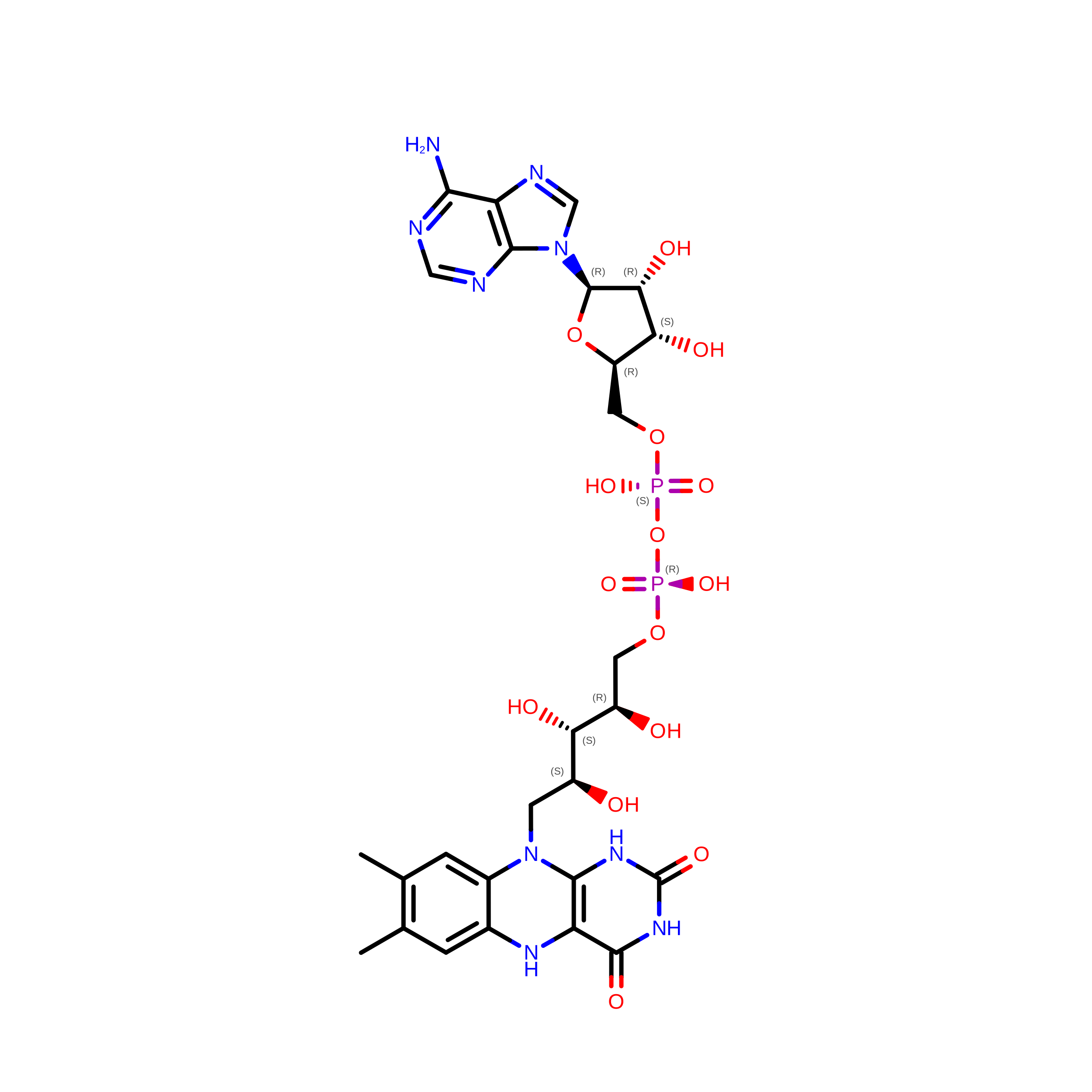
    The metazoan tRNA ligase complex (tRNA-LC) has essential roles in tRNA biogenesis and unfolded protein response. Its catalytic subunit RTCB contains a conserved active-site cysteine that is susceptible to metal ion-induced oxidative inactivation. The flavin-containing oxidoreductase PYROXD1 preserves the activity of human tRNA-LC in a NAD(P)H-dependent manner, but its protective mechanism remains elusive. Here, we report a cryogenic electron microscopic structure of the human RTCB-PYROXD1 complex, revealing that PYROXD1 directly interacts with the catalytic center of RTCB through its carboxy-terminal tail. NAD(P)H binding and FAD reduction allosterically control PYROXD1 activity and RTCB recruitment, while reoxidation of PYROXD1 enables timed release of RTCB. PYROXD1 interaction is mutually exclusive with Archease-mediated RTCB guanylylation, and guanylylated RTCB is intrinsically protected from oxidative inactivation. Together, these findings provide a mechanistic framework for the protective function of PYROXD1 that maintains the activity of the tRNA-LC under aerobic conditions.


  • Organizational Affiliation: 
    • Department of Biochemistry, University of Zurich, Zurich, Switzerland.

Macromolecule Content 

  • Total Structure Weight: 113.07 kDa 
  • Atom Count: 6,930 
  • Modeled Residue Count: 882 
  • Deposited Residue Count: 1,010 
  • Unique protein chains: 2

Macromolecules

Find similar proteins by:|  3D Structure
Entity ID: 1
MoleculeChains  Sequence LengthOrganismDetailsImage
RNA-splicing ligase RtcB homolog508Homo sapiensMutation(s): 0 
Gene Names: RTCB, C22orf28, HSPC117
EC: 6.5.1.8
UniProt & NIH Common Fund Data Resources
Find proteins for Q9Y3I0 (Homo sapiens)
Explore Q9Y3I0 
Go to UniProtKB:  Q9Y3I0
PHAROS:  Q9Y3I0
GTEx:  ENSG00000100220 
Entity Groups
Sequence Clusters30% Identity50% Identity70% Identity90% Identity95% Identity100% Identity
UniProt GroupQ9Y3I0
Sequence Annotations
Expand
Reference Sequence
Find similar proteins by:|  3D Structure
Entity ID: 2
MoleculeChains  Sequence LengthOrganismDetailsImage
Pyridine nucleotide-disulfide oxidoreductase domain-containing protein 1502Homo sapiensMutation(s): 0 
Gene Names: PYROXD1
EC: 1.8.1 (PDB Primary Data), 1.6.99.1 (UniProt)
UniProt & NIH Common Fund Data Resources
Find proteins for Q8WU10 (Homo sapiens)
Explore Q8WU10 
Go to UniProtKB:  Q8WU10
PHAROS:  Q8WU10
GTEx:  ENSG00000121350 
Entity Groups
Sequence Clusters30% Identity50% Identity70% Identity90% Identity95% Identity100% Identity
UniProt GroupQ8WU10
Sequence Annotations
Expand
Reference Sequence

Experimental Data & Validation

Experimental Data

  • Method: ELECTRON MICROSCOPY
  • Resolution: 3.30 Å
  • Aggregation State: PARTICLE 
  • Reconstruction Method: SINGLE PARTICLE 
EM Software:
TaskSoftware PackageVersion
MODEL REFINEMENTPHENIX
RECONSTRUCTIONcryoSPARC3.3.2

Structure Validation

View Full Validation Report


Entry History 

& Funding Information

Deposition Data


Funding OrganizationLocationGrant Number
Swiss National Science FoundationSwitzerland31003A_182567

Revision History  (Full details and data files)

  • Version 1.0: 2024-10-30
    Type: Initial release
  • Version 1.1: 2025-05-07
    Changes: Data collection, Database references