8OQ3 | pdb_00008oq3

Structure of methylamine treated human complement C3


Experimental Data Snapshot

  • Method: ELECTRON MICROSCOPY
  • Resolution: 2.90 Å
  • Aggregation State: PARTICLE 
  • Reconstruction Method: SINGLE PARTICLE 

wwPDB Validation 3D Report Full Report

Validation slider image for 8OQ3

This is version 1.3 of the entry. See complete history

Literature

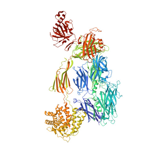
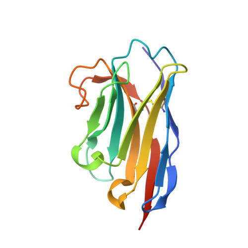
Cryo-EM analysis of complement C3 reveals a reversible major opening of the macroglobulin ring.

Gadeberg, T.A.F.Jorgensen, M.H.Olesen, H.G.Lorentzen, J.Harwood, S.L.Almeida, A.V.Fruergaard, M.U.Jensen, R.K.Kanis, P.Pedersen, H.Tranchant, E.Petersen, S.V.Thogersen, I.B.Kragelund, B.B.Lyons, J.A.Enghild, J.J.Andersen, G.R.

(2025) Nat Struct Mol Biol 

  • DOI: https://doi.org/10.1038/s41594-024-01467-4
  • Primary Citation Related Structures: 
    8OQ3

  • PubMed Abstract: 

    The C3 protein is the central molecule within the complement system and undergoes proteolytic activation to C3b in the presence of pathogens. Pattern-independent activation of C3 also occurs via hydrolysis, resulting in C3(H 2 O), but the structural details of C3 hydrolysis remain elusive. Here we show that the conformation of the C3(H 2 O) analog, C3MA, is indistinguishable from C3b. In contrast, the reaction intermediate C3* adopts a conformation dramatically different from both C3 and C3MA. In C3*, unlocking of the macroglobulin (MG) 3 domain creates a large opening in the MG ring through which the anaphylatoxin (ANA) domain translocates through a transient opening. C3MA formation is inhibited by an MG3-specific nanobody and prevented by linking the ANA domain to the C3 β-chain. Our study reveals an unexpected dynamic behavior of C3 and forms the basis for elucidation of the in vivo contribution of C3 hydrolysis and for controlling complement upon intravascular hemolysis and surface-contact-induced activation.


  • Organizational Affiliation
    • Department of Molecular Biology and Genetics, Aarhus, Denmark.

Macromolecule Content 

  • Total Structure Weight: 400.37 kDa 
  • Atom Count: 26,596 
  • Modeled Residue Count: 3,358 
  • Deposited Residue Count: 3,540 
  • Unique protein chains: 2

Macromolecules

Find similar proteins by:|  3D Structure
Entity ID: 1
MoleculeChains  Sequence LengthOrganismDetailsImage
Complement C3A,
C [auth D]
1,641Homo sapiensMutation(s): 0 
UniProt & NIH Common Fund Data Resources
Find proteins for P01024 (Homo sapiens)
Explore P01024 
Go to UniProtKB:  P01024
PHAROS:  P01024
GTEx:  ENSG00000125730 
Entity Groups
Sequence Clusters30% Identity50% Identity70% Identity90% Identity95% Identity100% Identity
UniProt GroupP01024
Glycosylation
Glycosylation Sites: 2Go to GlyGen: P01024-1
Sequence Annotations
Expand
Reference Sequence
Find similar proteins by:|  3D Structure
Entity ID: 2
MoleculeChains  Sequence LengthOrganismDetailsImage
nanobody hC3Nb1 with mutationB [auth C],
D [auth F]
129Lama glamaMutation(s): 0 

Oligosaccharides

Help  
Entity ID: 3
MoleculeChains Length2D Diagram GlycosylationD Interactions
beta-D-mannopyranose-(1-4)-2-acetamido-2-deoxy-beta-D-glucopyranose-(1-4)-2-acetamido-2-deoxy-beta-D-glucopyranoseE [auth G],
F [auth H]
3N-Glycosylation
Glycosylation Resources
GlyTouCan: G15407YE
GlyCosmos: G15407YE
GlyGen: G15407YE

Experimental Data & Validation

Experimental Data

  • Method: ELECTRON MICROSCOPY
  • Resolution: 2.90 Å
  • Aggregation State: PARTICLE 
  • Reconstruction Method: SINGLE PARTICLE 
EM Software:
TaskSoftware PackageVersion
MODEL REFINEMENTPHENIX

Structure Validation

View Full Validation Report


Entry History 

& Funding Information

Deposition Data


Funding OrganizationLocationGrant Number
LundbeckfondenDenmarkR155-2015-2666
Novo Nordisk FoundationDenmarkNNF18OC0052105
Danish Council for Independent ResearchDenmark4181-00137B

Revision History  (Full details and data files)

  • Version 1.0: 2024-02-21
    Type: Initial release
  • Version 1.1: 2024-10-23
    Changes: Data collection, Structure summary
  • Version 1.2: 2025-03-05
    Changes: Data collection, Database references
  • Version 1.3: 2025-07-02
    Changes: Data collection