8ONH

Variant Surface Glycoprotein VSG11wt-Oil


Experimental Data Snapshot

  • Method: X-RAY DIFFRACTION
  • Resolution: 2.59 Å
  • R-Value Free: 0.270 
  • R-Value Work: 0.229 
  • R-Value Observed: 0.231 

wwPDB Validation   3D Report Full Report


Ligand Structure Quality Assessment 


This is version 1.0 of the entry. See complete history


Literature

A structural classification of the variant surface glycoproteins of the African trypanosomey.

Dakovic, S.Zeelen, J.P.Gkeka, A.Chandra, M.van Straaten, M.Foti, K.Zhong, J.Vlachou, E.P.Aresta-Branco, F.Verdi, J.P.Papavasiliou, F.N.Stebbins, C.E.

(2023) PLoS Negl Trop Dis 17: e0011621-e0011621

  • DOI: https://doi.org/10.1371/journal.pntd.0011621
  • Primary Citation of Related Structures:  
    8OK4, 8OK5, 8OK6, 8OK7, 8OK8, 8ONH, 8Q0E, 8Q0P

  • PubMed Abstract: 

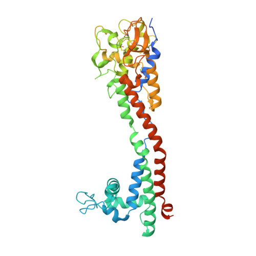
    Long-term immune evasion by the African trypanosome is achieved through repetitive cycles of surface protein replacement with antigenically distinct versions of the dense Variant Surface Glycoprotein (VSG) coat. Thousands of VSG genes and pseudo-genes exist in the parasite genome that, together with genetic recombination mechanisms, allow for essentially unlimited immune escape from the adaptive immune system of the host. The diversity space of the "VSGnome" at the protein level was thought to be limited to a few related folds whose structures were determined more than 30 years ago. However, recent progress has shown that the VSGs possess significantly more architectural variation than had been appreciated. Here we combine experimental X-ray crystallography (presenting structures of N-terminal domains of coat proteins VSG11, VSG21, VSG545, VSG558, and VSG615) with deep-learning prediction using Alphafold to produce models of hundreds of VSG proteins. We classify the VSGnome into groups based on protein architecture and oligomerization state, contextualize recent bioinformatics clustering schemes, and extensively map VSG-diversity space. We demonstrate that in addition to the structural variability and post-translational modifications observed thus far, VSGs are also characterized by variations in oligomerization state and possess inherent flexibility and alternative conformations, lending additional variability to what is exposed to the immune system. Finally, these additional experimental structures and the hundreds of Alphafold predictions confirm that the molecular surfaces of the VSGs remain distinct from variant to variant, supporting the hypothesis that protein surface diversity is central to the process of antigenic variation used by this organism during infection.


  • Organizational Affiliation

    Division of Structural Biology of Infection and Immunity, German Cancer Research Center, Heidelberg, Germany.


Macromolecules
Find similar proteins by:  (by identity cutoff)  |  3D Structure
Entity ID: 1
MoleculeChains Sequence LengthOrganismDetailsImage
Variant surface glycoprotein
A, B, C, D, E
A, B, C, D, E, F, G, H, I, J, K, L, M, N, O, P, Q, R
410Trypanosoma brucei bruceiMutation(s): 0 
Gene Names: Tb427.BES126.15
UniProt
Find proteins for B3GVS7 (Trypanosoma brucei brucei)
Explore B3GVS7 
Go to UniProtKB:  B3GVS7
Entity Groups  
Sequence Clusters30% Identity50% Identity70% Identity90% Identity95% Identity100% Identity
UniProt GroupB3GVS7
Sequence Annotations
Expand
  • Reference Sequence
Oligosaccharides

Help

Entity ID: 2
MoleculeChains Length2D Diagram Glycosylation3D Interactions
alpha-D-mannopyranose-(1-2)-alpha-D-mannopyranose-(1-3)-[alpha-D-mannopyranose-(1-6)]beta-D-mannopyranose-(1-4)-2-acetamido-2-deoxy-beta-D-glucopyranose-(1-4)-2-acetamido-2-deoxy-beta-D-glucopyranose
S, Z
6N-Glycosylation
Glycosylation Resources
GlyTouCan:  G56014GC
GlyCosmos:  G56014GC
GlyGen:  G56014GC
Entity ID: 3
MoleculeChains Length2D Diagram Glycosylation3D Interactions
alpha-D-mannopyranose-(1-3)-[alpha-D-mannopyranose-(1-6)]beta-D-mannopyranose-(1-4)-2-acetamido-2-deoxy-beta-D-glucopyranose-(1-3)-2-acetamido-2-deoxy-beta-D-glucopyranose
T
5N-Glycosylation
Glycosylation Resources
GlyTouCan:  G32708SB
GlyCosmos:  G32708SB
GlyGen:  G32708SB
Entity ID: 4
MoleculeChains Length2D Diagram Glycosylation3D Interactions
beta-D-mannopyranose-(1-4)-2-acetamido-2-deoxy-beta-D-glucopyranose-(1-3)-2-acetamido-2-deoxy-beta-D-glucopyranose
U
3N-Glycosylation
Glycosylation Resources
GlyTouCan:  G53081KU
GlyCosmos:  G53081KU
GlyGen:  G53081KU
Entity ID: 5
MoleculeChains Length2D Diagram Glycosylation3D Interactions
beta-D-mannopyranose-(1-4)-2-acetamido-2-deoxy-beta-D-glucopyranose-(1-4)-2-acetamido-2-deoxy-beta-D-glucopyranose
V, X, Y
3N-Glycosylation
Glycosylation Resources
GlyTouCan:  G15407YE
GlyCosmos:  G15407YE
GlyGen:  G15407YE
Entity ID: 6
MoleculeChains Length2D Diagram Glycosylation3D Interactions
alpha-D-mannopyranose-(1-3)-[alpha-D-mannopyranose-(1-6)]beta-D-mannopyranose-(1-3)-2-acetamido-2-deoxy-beta-D-glucopyranose-(1-3)-2-acetamido-2-deoxy-beta-D-glucopyranoseCA [auth c],
W
5N-Glycosylation
Entity ID: 7
MoleculeChains Length2D Diagram Glycosylation3D Interactions
alpha-D-mannopyranose-(1-3)-[alpha-D-mannopyranose-(1-6)]beta-D-mannopyranose-(1-4)-2-acetamido-2-deoxy-beta-D-glucopyranose-(1-4)-2-acetamido-2-deoxy-beta-D-glucopyranoseAA [auth a],
BA [auth b]
5N-Glycosylation
Glycosylation Resources
GlyTouCan:  G22768VO
GlyCosmos:  G22768VO
GlyGen:  G22768VO
Small Molecules
Ligands 2 Unique
IDChains Name / Formula / InChI Key2D Diagram3D Interactions
NAG (Subject of Investigation/LOI)
Query on NAG

Download Ideal Coordinates CCD File 
IA [auth E],
SA [auth O],
UA [auth Q],
WA [auth R]
2-acetamido-2-deoxy-beta-D-glucopyranose
C8 H15 N O6
OVRNDRQMDRJTHS-FMDGEEDCSA-N
GLC (Subject of Investigation/LOI)
Query on GLC

Download Ideal Coordinates CCD File 
DA [auth A]
EA [auth B]
FA [auth C]
GA [auth D]
HA [auth E]
DA [auth A],
EA [auth B],
FA [auth C],
GA [auth D],
HA [auth E],
JA [auth F],
KA [auth H],
LA [auth I],
MA [auth J],
NA [auth K],
OA [auth L],
PA [auth M],
QA [auth N],
RA [auth O],
TA [auth P],
VA [auth Q],
XA [auth R]
alpha-D-glucopyranose
C6 H12 O6
WQZGKKKJIJFFOK-DVKNGEFBSA-N
Experimental Data & Validation

Experimental Data

  • Method: X-RAY DIFFRACTION
  • Resolution: 2.59 Å
  • R-Value Free: 0.270 
  • R-Value Work: 0.229 
  • R-Value Observed: 0.231 
  • Space Group: P 1 21 1
Unit Cell:
Length ( Å )Angle ( ˚ )
a = 131.88α = 90
b = 210.771β = 104.537
c = 133.754γ = 90
Software Package:
Software NamePurpose
PHENIXrefinement
xia2data reduction
Aimlessdata scaling
PHENIXphasing
Cootmodel building

Structure Validation

View Full Validation Report


Ligand Structure Quality Assessment 


Entry History & Funding Information

Deposition Data


Funding OrganizationLocationGrant Number
Not funded--

Revision History  (Full details and data files)

  • Version 1.0: 2023-09-13
    Type: Initial release