8OLC | pdb_00008olc

SA11 Rotavirus Trypsinized Triple Layered Particle


Experimental Data Snapshot

  • Method: ELECTRON MICROSCOPY
  • Resolution: 3.48 Å
  • Aggregation State: PARTICLE 
  • Reconstruction Method: SINGLE PARTICLE 

wwPDB Validation 3D Report Full Report

Validation slider image for 8OLC

This is version 1.2 of the entry. See complete history

Literature

Structural determinants of rotavirus proteolytic activation.

Asensio-Cob, D.Mata, C.P.Gomez-Blanco, J.Vargas, J.Rodriguez, J.M.Luque, D.

(2025) PLoS Pathog 21: e1013063-e1013063

  • DOI: https://doi.org/10.1371/journal.ppat.1013063
  • Primary Citation Related Structures: 
    8OLB, 8OLC, 8OLE, 8QTZ

  • PubMed Abstract: 

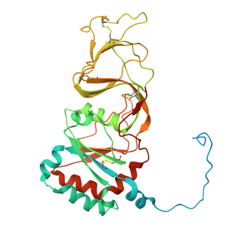
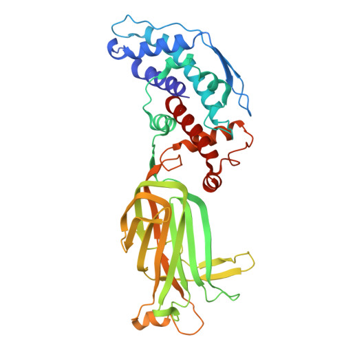
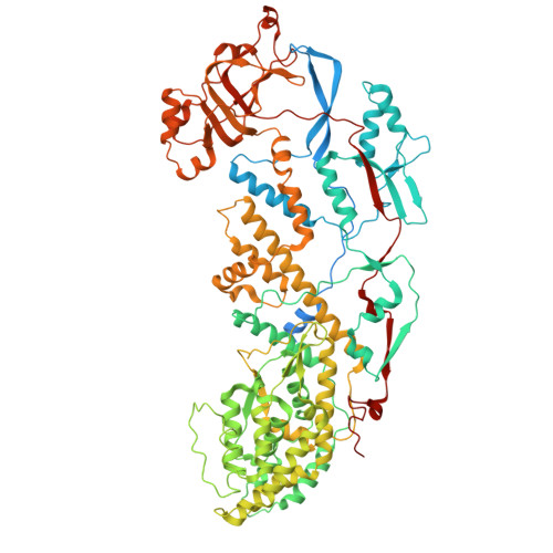
    The infectivity of rotavirus (RV), the leading cause of childhood diarrhea, hinges on the activation of viral particles through the proteolysis of the spike protein by trypsin-like proteases in the host intestinal lumen. In order to determine the structural basis of trypsin activation, we have used cryogenic electron microscopy (cryo-EM) and advanced image processing methods to compare uncleaved and cleaved RV particles. We find that the conformation of the non-proteolyzed spike is constrained by the position of loops that surround its structure, linking the lectin domains of the spike head to its body. The proteolysis of these loops removes this structural constraint, thereby enabling the spike to undergo the necessary conformational changes required for cell membrane penetration. Thus, these loops function as regulatory elements to ensure that the spike protein is activated precisely when and where it is needed to facilitate a successful infection.


  • Organizational Affiliation
    • Department of Molecular Medicine, Peter Gilgan Centre for Research and Learning, The Hospital for Sick Children, Toronto, Ontario, Canada.

Macromolecule Content 

  • Total Structure Weight: 1,277.02 kDa 
  • Atom Count: 82,143 
  • Modeled Residue Count: 10,266 
  • Deposited Residue Count: 11,163 
  • Unique protein chains: 3

Macromolecules

Find similar proteins by:|  3D Structure
Entity ID: 1
MoleculeChains  Sequence LengthOrganismDetailsImage
Outer capsid glycoprotein VP7326RotavirusMutation(s): 0 
Gene Names: VP7VP1
UniProt
Find proteins for A0A060IEQ1 (Rotavirus A)
Explore A0A060IEQ1 
Go to UniProtKB:  A0A060IEQ1
Entity Groups
Sequence Clusters30% Identity50% Identity70% Identity90% Identity95% Identity100% Identity
UniProt GroupA0A060IEQ1
Glycosylation
Glycosylation Sites: 1
Sequence Annotations
Expand
Reference Sequence
Find similar proteins by:|  3D Structure
Entity ID: 2
MoleculeChains  Sequence LengthOrganismDetailsImage
Intermediate capsid protein VP6397RotavirusMutation(s): 0 
Gene Names: VP6
UniProt
Find proteins for A2T3S6 (Rotavirus A)
Explore A2T3S6 
Go to UniProtKB:  A2T3S6
Entity Groups
Sequence Clusters30% Identity50% Identity70% Identity90% Identity95% Identity100% Identity
UniProt GroupA2T3S6
Sequence Annotations
Expand
Reference Sequence
Find similar proteins by:|  3D Structure
Entity ID: 3
MoleculeChains  Sequence LengthOrganismDetailsImage
Inner capsid protein VP2AA [auth A],
BA [auth B]
882RotavirusMutation(s): 0 
Gene Names: VP2
UniProt
Find proteins for A2T3R1 (Rotavirus A)
Explore A2T3R1 
Go to UniProtKB:  A2T3R1
Entity Groups
Sequence Clusters30% Identity50% Identity70% Identity90% Identity95% Identity100% Identity
UniProt GroupA2T3R1
Sequence Annotations
Expand
Reference Sequence

Small Molecules

Ligands 3 Unique
IDChains Name / Formula / InChI Key2D Diagram3D Interactions
NAG
(Subject of Investigation/LOI)

Query on NAG


Download:Ideal Coordinates CCD File
AB [auth l]
DA [auth d]
DB [auth m]
GB [auth n]
HA [auth e]
AB [auth l],
DA [auth d],
DB [auth m],
GB [auth n],
HA [auth e],
JB [auth o],
KA [auth f],
PA [auth h],
UA [auth j],
XA [auth k]
2-acetamido-2-deoxy-beta-D-glucopyranose
C8 H15 N O6
OVRNDRQMDRJTHS-FMDGEEDCSA-N
ZN
(Subject of Investigation/LOI)

Query on ZN


Download:Ideal Coordinates CCD File
MB [auth C],
NB [auth F],
OB [auth I],
PB [auth L],
QB [auth O]
ZINC ION
Zn
PTFCDOFLOPIGGS-UHFFFAOYSA-N
CA
(Subject of Investigation/LOI)

Query on CA


Download:Ideal Coordinates CCD File
BB [auth l]
CA [auth c]
CB [auth l]
EA [auth d]
EB [auth m]
BB [auth l],
CA [auth c],
CB [auth l],
EA [auth d],
EB [auth m],
FA [auth d],
FB [auth m],
GA [auth d],
HB [auth n],
IA [auth e],
IB [auth n],
JA [auth e],
KB [auth o],
LA [auth f],
LB [auth o],
MA [auth f],
NA [auth g],
OA [auth g],
QA [auth h],
RA [auth h],
SA [auth i],
TA [auth i],
VA [auth j],
WA [auth j],
YA [auth k],
ZA [auth k]
CALCIUM ION
Ca
BHPQYMZQTOCNFJ-UHFFFAOYSA-N

Experimental Data & Validation

Experimental Data

  • Method: ELECTRON MICROSCOPY
  • Resolution: 3.48 Å
  • Aggregation State: PARTICLE 
  • Reconstruction Method: SINGLE PARTICLE 
EM Software:
TaskSoftware PackageVersion
RECONSTRUCTIONRELION

Structure Validation

View Full Validation Report


Entry History 

& Funding Information

Deposition Data


Funding OrganizationLocationGrant Number
Spanish Ministry of Science, Innovation, and UniversitiesSpainISCIII-AESI PI20CIII/00014

Revision History  (Full details and data files)

  • Version 1.0: 2024-09-25
    Type: Initial release
  • Version 1.1: 2024-11-13
    Changes: Data collection, Structure summary
  • Version 1.2: 2025-10-08
    Changes: Data collection, Database references