8OIZ

Crystal structure of human CRBN-DDB1 in complex with Pomalidomide


Experimental Data Snapshot

  • Method: X-RAY DIFFRACTION
  • Resolution: 2.50 Å
  • R-Value Free: 0.262 
  • R-Value Work: 0.213 
  • R-Value Observed: 0.216 

wwPDB Validation   3D Report Full Report


Ligand Structure Quality Assessment 


This is version 1.1 of the entry. See complete history


Literature

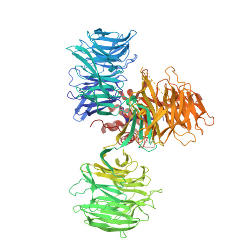
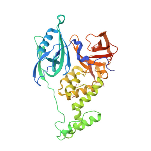
A Degron Blocking Strategy Towards Improved CRL4 CRBN Recruiting PROTAC Selectivity.

Bouguenina, H.Scarpino, A.O'Hanlon, J.A.Warne, J.Wang, H.Z.Wah Hak, L.C.Sadok, A.McAndrew, P.C.Stubbs, M.Pierrat, O.A.Hahner, T.Cabry, M.P.Le Bihan, Y.V.Mitsopoulos, C.Sialana, F.J.Roumeliotis, T.I.Burke, R.van Montfort, R.L.M.Choudhari, J.Chopra, R.Caldwell, J.J.Collins, I.

(2023) Chembiochem 24: e202300351-e202300351

  • DOI: https://doi.org/10.1002/cbic.202300351
  • Primary Citation of Related Structures:  
    8OIZ, 8OJH

  • PubMed Abstract: 

    Small molecules inducing protein degradation are important pharmacological tools to interrogate complex biology and are rapidly translating into clinical agents. However, to fully realise the potential of these molecules, selectivity remains a limiting challenge. Herein, we addressed the issue of selectivity in the design of CRL4 CRBN recruiting PROteolysis TArgeting Chimeras (PROTACs). Thalidomide derivatives used to generate CRL4 CRBN recruiting PROTACs have well described intrinsic monovalent degradation profiles by inducing the recruitment of neo-substrates, such as GSPT1, Ikaros and Aiolos. We leveraged structural insights from known CRL4 CRBN neo-substrates to attenuate and indeed remove this monovalent degradation function in well-known CRL4 CRBN molecular glues degraders, namely CC-885 and Pomalidomide. We then applied these design principles on a previously published BRD9 PROTAC (dBRD9-A) and generated an analogue with improved selectivity profile. Finally, we implemented a computational modelling pipeline to show that our degron blocking design does not impact PROTAC-induced ternary complex formation. We believe that the tools and principles presented in this work will be valuable to support the development of targeted protein degradation.


  • Organizational Affiliation

    Centre for Cancer Drug Discovery, Institute of Cancer Research, 15 Cotswold Road, Sutton, London, SM2 5NG, UK.


Macromolecules
Find similar proteins by:  (by identity cutoff)  |  3D Structure
Entity ID: 1
MoleculeChains Sequence LengthOrganismDetailsImage
DNA damage-binding protein 11,148Homo sapiensMutation(s): 0 
Gene Names: DDB1XAP1
UniProt & NIH Common Fund Data Resources
Find proteins for Q16531 (Homo sapiens)
Explore Q16531 
Go to UniProtKB:  Q16531
PHAROS:  Q16531
GTEx:  ENSG00000167986 
Entity Groups  
Sequence Clusters30% Identity50% Identity70% Identity90% Identity95% Identity100% Identity
UniProt GroupQ16531
Sequence Annotations
Expand
  • Reference Sequence
Find similar proteins by:  (by identity cutoff)  |  3D Structure
Entity ID: 2
MoleculeChains Sequence LengthOrganismDetailsImage
Protein cereblon407Homo sapiensMutation(s): 0 
Gene Names: CRBNAD-006
UniProt & NIH Common Fund Data Resources
Find proteins for Q96SW2 (Homo sapiens)
Explore Q96SW2 
Go to UniProtKB:  Q96SW2
PHAROS:  Q96SW2
GTEx:  ENSG00000113851 
Entity Groups  
Sequence Clusters30% Identity50% Identity70% Identity90% Identity95% Identity100% Identity
UniProt GroupQ96SW2
Sequence Annotations
Expand
  • Reference Sequence
Small Molecules
Ligands 3 Unique
IDChains Name / Formula / InChI Key2D Diagram3D Interactions
Y70 (Subject of Investigation/LOI)
Query on Y70

Download Ideal Coordinates CCD File 
J [auth B]S-Pomalidomide
C13 H11 N3 O4
UVSMNLNDYGZFPF-QMMMGPOBSA-N
ZN
Query on ZN

Download Ideal Coordinates CCD File 
I [auth B]ZINC ION
Zn
PTFCDOFLOPIGGS-UHFFFAOYSA-N
EDO
Query on EDO

Download Ideal Coordinates CCD File 
C [auth A]
D [auth A]
E [auth A]
F [auth A]
G [auth A]
C [auth A],
D [auth A],
E [auth A],
F [auth A],
G [auth A],
H [auth A]
1,2-ETHANEDIOL
C2 H6 O2
LYCAIKOWRPUZTN-UHFFFAOYSA-N
Experimental Data & Validation

Experimental Data

  • Method: X-RAY DIFFRACTION
  • Resolution: 2.50 Å
  • R-Value Free: 0.262 
  • R-Value Work: 0.213 
  • R-Value Observed: 0.216 
  • Space Group: P 21 21 21
Unit Cell:
Length ( Å )Angle ( ˚ )
a = 71.841α = 90
b = 128.508β = 90
c = 198.377γ = 90
Software Package:
Software NamePurpose
BUSTERrefinement
Aimlessdata scaling
PHASERphasing
PDB_EXTRACTdata extraction
DIALSdata reduction

Structure Validation

View Full Validation Report


Ligand Structure Quality Assessment 


Entry History & Funding Information

Deposition Data


Funding OrganizationLocationGrant Number
Cancer Research UKUnited KingdomC309/A11566

Revision History  (Full details and data files)

  • Version 1.0: 2023-07-19
    Type: Initial release
  • Version 1.1: 2023-12-13
    Changes: Data collection, Database references