8OIG | pdb_00008oig

Crystal Structure of Staphopain C from Staphylococcus aureus


Experimental Data Snapshot

  • Method: X-RAY DIFFRACTION
  • Resolution: 1.58 Å
  • R-Value Free: 
    0.116 (Depositor), 0.138 (DCC) 
  • R-Value Work: 
    0.088 (Depositor), 0.110 (DCC) 
  • R-Value Observed: 
    0.088 (Depositor) 

Starting Model: in silico
View more details

wwPDB Validation 3D Report Full Report

Validation slider image for 8OIG

Ligand Structure Quality Assessment 


This is version 1.1 of the entry. See complete history

Literature

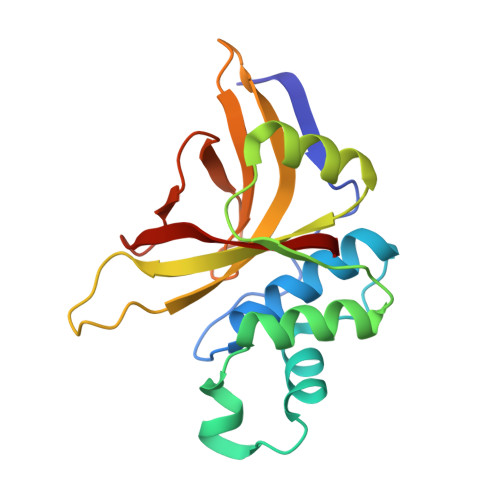
Crystal Structure of Staphopain C from Staphylococcus aureus.

Magoch, M.McEwen, A.G.Napolitano, V.Wladyka, B.Dubin, G.

(2023) Molecules 28

  • DOI: https://doi.org/10.3390/molecules28114407
  • Primary Citation Related Structures: 
    8OIG

  • PubMed Abstract: 

    Staphylococcus aureus is a common opportunistic pathogen of humans and livestock that causes a wide variety of infections. The success of S. aureus as a pathogen depends on the production of an array of virulence factors including cysteine proteases (staphopains)-major secreted proteases of certain strains of the bacterium. Here, we report the three-dimensional structure of staphopain C (ScpA2) of S. aureus , which shows the typical papain-like fold and uncovers a detailed molecular description of the active site. Because the protein is involved in the pathogenesis of a chicken disease, our work provides the foundation for inhibitor design and potential antimicrobial strategies against this pathogen.


  • Organizational Affiliation
    • Malopolska Centre of Biotechnology, Jagiellonian University, 30-387 Krakow, Poland.

Macromolecule Content 

  • Total Structure Weight: 59.46 kDa 
  • Atom Count: 4,637 
  • Modeled Residue Count: 519 
  • Deposited Residue Count: 519 
  • Unique protein chains: 1

Macromolecules

Find similar proteins by:|  3D Structure
Entity ID: 1
MoleculeChains  Sequence LengthOrganismDetailsImage
Thiol protease
A, B, C
173Staphylococcus aureusMutation(s): 0 
UniProt
Find proteins for Q8RJP3 (Staphylococcus aureus)
Explore Q8RJP3 
Go to UniProtKB:  Q8RJP3
Entity Groups
Sequence Clusters30% Identity50% Identity70% Identity90% Identity95% Identity100% Identity
UniProt GroupQ8RJP3
Sequence Annotations
Expand
Reference Sequence

Small Molecules

Ligands 3 Unique
IDChains Name / Formula / InChI Key2D Diagram3D Interactions
GOL
(Subject of Investigation/LOI)

Query on GOL


Download:Ideal Coordinates CCD File
D [auth A],
E [auth A],
L [auth C]
GLYCEROL
C3 H8 O3
PEDCQBHIVMGVHV-UHFFFAOYSA-N
EDO
(Subject of Investigation/LOI)

Query on EDO


Download:Ideal Coordinates CCD File
M [auth C]1,2-ETHANEDIOL
C2 H6 O2
LYCAIKOWRPUZTN-UHFFFAOYSA-N
CL
(Subject of Investigation/LOI)

Query on CL


Download:Ideal Coordinates CCD File
F [auth B]
G [auth B]
H [auth C]
I [auth C]
J [auth C]
F [auth B],
G [auth B],
H [auth C],
I [auth C],
J [auth C],
K [auth C]
CHLORIDE ION
Cl
VEXZGXHMUGYJMC-UHFFFAOYSA-M

Experimental Data & Validation

Experimental Data

  • Method: X-RAY DIFFRACTION
  • Resolution: 1.58 Å
  • R-Value Free:  0.116 (Depositor), 0.138 (DCC) 
  • R-Value Work:  0.088 (Depositor), 0.110 (DCC) 
  • R-Value Observed: 0.088 (Depositor) 
Space Group: P 31
Unit Cell:
Length ( Å )Angle ( ˚ )
a = 103.136α = 90
b = 103.136β = 90
c = 51.204γ = 120
Software Package:
Software NamePurpose
REFMACrefinement
AutoProcessdata reduction
AutoProcessdata scaling
PHASERphasing

Structure Validation

View Full Validation Report


Ligand Structure Quality Assessment 


Entry History 

& Funding Information

Deposition Data


Funding OrganizationLocationGrant Number
Polish National Science CentrePolandUMO-2011/01/D/NZ1/01169
Polish National Science CentrePolandN N303 813340

Revision History  (Full details and data files)

  • Version 1.0: 2023-06-21
    Type: Initial release
  • Version 1.1: 2024-06-19
    Changes: Data collection