8OID | pdb_00008oid

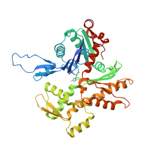
Cryo-EM structure of ADP-bound, filamentous beta-actin harboring the N111S mutation


Experimental Data Snapshot

  • Method: ELECTRON MICROSCOPY
  • Resolution: 2.30 Å
  • Aggregation State: FILAMENT 
  • Reconstruction Method: SINGLE PARTICLE 

Starting Model: experimental
View more details

wwPDB Validation 3D Report Full Report

Validation slider image for 8OID

This is version 1.4 of the entry. See complete history. 

Literature

Molecular mechanisms of inorganic-phosphate release from the core and barbed end of actin filaments.

Oosterheert, W., Blanc, F.E.C., Roy, A., Belyy, A., Sanders, M.B., Hofnagel, O., Hummer, G., Bieling, P., Raunser, S.

(2023) Nat Struct Mol Biol 30: 1774-1785

  • DOI: https://doi.org/10.1038/s41594-023-01101-9
  • Primary Citation Related Structures: 
    8OI6, 8OI8, 8OID

  • PubMed Abstract: 

    The release of inorganic phosphate (P i ) from actin filaments constitutes a key step in their regulated turnover, which is fundamental to many cellular functions. The mechanisms underlying P i release from the core and barbed end of actin filaments remain unclear. Here, using human and bovine actin isoforms, we combine cryo-EM with molecular-dynamics simulations and in vitro reconstitution to demonstrate how actin releases P i through a 'molecular backdoor'. While constantly open at the barbed end, the backdoor is predominantly closed in filament-core subunits and opens only transiently through concerted amino acid rearrangements. This explains why P i escapes rapidly from the filament end but slowly from internal subunits. In a nemaline-myopathy-associated actin variant, the backdoor is predominantly open in filament-core subunits, resulting in accelerated P i release and filaments with drastically shortened ADP-P i caps. Our results provide the molecular basis for P i release from actin and exemplify how a disease-linked mutation distorts the nucleotide-state distribution and atomic structure of the filament.


  • Organizational Affiliation: 
    • Department of Structural Biochemistry, Max Planck Institute of Molecular Physiology, Dortmund, Germany.

Macromolecule Content 

  • Total Structure Weight: 210.94 kDa 
  • Atom Count: 15,206 
  • Modeled Residue Count: 1,850 
  • Deposited Residue Count: 1,875 
  • Unique protein chains: 1

Macromolecules

Find similar proteins by:|  3D Structure
Entity ID: 1
MoleculeChains  Sequence LengthOrganismDetailsImage
Actin, cytoplasmic 1, N-terminally processedA [auth C],
B [auth A],
C [auth B],
D,
E
375Homo sapiensMutation(s): 2 
Gene Names: ACTB
EC: 3.6.4
UniProt & NIH Common Fund Data Resources
Find proteins for P60709 (Homo sapiens)
Explore P60709 
Go to UniProtKB:  P60709
PHAROS:  P60709
GTEx:  ENSG00000075624 
Entity Groups
Sequence Clusters30% Identity50% Identity70% Identity90% Identity95% Identity100% Identity
UniProt GroupP60709
Sequence Annotations
Expand
Reference Sequence

Small Molecules

Ligands 2 Unique
IDChains Name / Formula / InChI Key2D Diagram3D Interactions
ADP
(Subject of Investigation/LOI)

Query on ADP


Download:Ideal Coordinates CCD File
F [auth C],
H [auth A],
J [auth B],
L [auth D],
N [auth E]
ADENOSINE-5'-DIPHOSPHATE
C10 H15 N5 O10 P2
XTWYTFMLZFPYCI-KQYNXXCUSA-N
MG
(Subject of Investigation/LOI)

Query on MG


Download:Ideal Coordinates CCD File
G [auth C],
I [auth A],
K [auth B],
M [auth D],
O [auth E]
MAGNESIUM ION
Mg
JLVVSXFLKOJNIY-UHFFFAOYSA-N
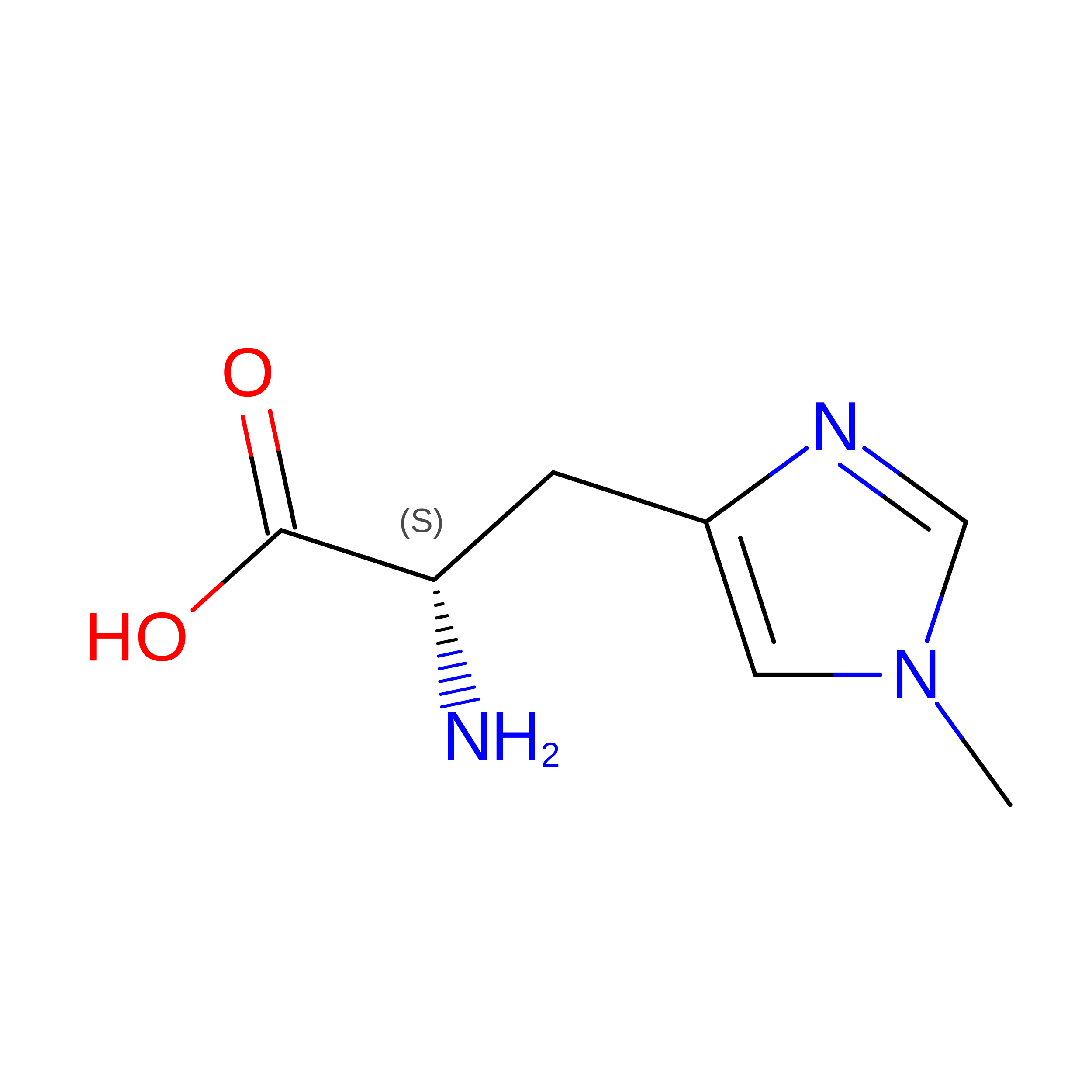
Modified Residues  1 Unique
IDChains TypeFormula2D DiagramParent
HIC
Query on HIC
A [auth C],
B [auth A],
C [auth B],
D,
E
L-PEPTIDE LINKINGC7 H11 N3 O2HIS

Experimental Data & Validation

Experimental Data

  • Method: ELECTRON MICROSCOPY
  • Resolution: 2.30 Å
  • Aggregation State: FILAMENT 
  • Reconstruction Method: SINGLE PARTICLE 
EM Software:
TaskSoftware PackageVersion
RECONSTRUCTIONRELION3.1.0
MODEL REFINEMENTPHENIX1.20.1-4487-000

Structure Validation

View Full Validation Report


Entry History 

& Funding Information

Deposition Data


Funding OrganizationLocationGrant Number
Max Planck SocietyGermany--
Alexander von Humboldt FoundationGermany--
European Research Council (ERC)European Union856118

Revision History  (Full details and data files)

  • Version 1.0: 2023-08-09
    Type: Initial release
  • Version 1.1: 2023-08-16
    Changes: Data collection, Database references, Refinement description, Source and taxonomy, Structure summary
  • Version 1.2: 2023-09-27
    Changes: Database references, Refinement description
  • Version 1.3: 2023-10-04
    Changes: Database references
  • Version 1.4: 2023-11-22
    Changes: Database references