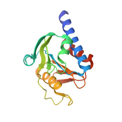
Crystallographic fragment screen of the c-di-AMP-synthesizing enzyme CdaA from Bacillus subtilis.
Garbers, T., Neumann, P., Wollenhaupt, J., Dickmanns, A., Weiss, M.S., Ficner, R.(2024) Acta Crystallogr F Struct Biol Commun 80: 200-209
- PubMed: 39177700 Search on PubMedSearch on PubMed Central
- DOI: https://doi.org/10.1107/S2053230X24007039
- Primary Citation Related Structures:
8OFH, 8OFO, 8OGM, 8OGN, 8OGO, 8OGP, 8OGQ, 8OGR, 8OGS, 8OGT, 8OGU, 8OGV, 8OGW, 8OGY, 8OGZ, 8OH0, 8OH1, 8OHB, 8OHC, 8OHE, 8OHF, 8OHG, 8OHH, 8OHJ, 8OHK, 8OHL, 8OHO, 9G0G - PubMed Abstract:
Crystallographic fragment screening has become a pivotal technique in structure-based drug design, particularly for bacterial targets with a crucial role in infectious disease mechanisms. The enzyme CdaA, which synthesizes an essential second messenger cyclic di-AMP (c-di-AMP) in many pathogenic bacteria, has emerged as a promising candidate for the development of novel antibiotics. To identify crystals suitable for fragment screening, CdaA enzymes from Streptococcus pneumoniae, Bacillus subtilis and Enterococcus faecium were purified and crystallized. Crystals of B. subtilis CdaA, which diffracted to the highest resolution of 1.1 Å, were used to perform the screening of 96 fragments, yielding data sets with resolutions spanning from 1.08 to 1.87 Å. A total of 24 structural hits across eight different sites were identified. Four fragments bind to regions that are highly conserved among pathogenic bacteria, specifically the active site (three fragments) and the dimerization interface (one fragment). The coordinates of the three active-site fragments were used to perform an in silico drug-repurposing screen using the OpenEye suite and the DrugBank database. This screen identified tenofovir, an approved drug, that is predicted to interact with the ATP-binding region of CdaA. Its inhibitory potential against pathogenic E. faecium CdaA has been confirmed by ITC measurements. These findings not only demonstrate the feasibility of this approach for identifying lead compounds for the design of novel antibacterial agents, but also pave the way for further fragment-based lead-optimization efforts targeting CdaA.
- Department of Molecular Structural Biology, Institute of Microbiology and Genetics, GZMB, Georg-August-University Göttingen, Justus-von-Liebig-Weg 11, 37073 Göttingen, Germany.
Organizational Affiliation: