8OG5

Crystal structure of human DCAF1 WD40 repeats (Q1250L) in complex with compound 1


Experimental Data Snapshot

  • Method: X-RAY DIFFRACTION
  • Resolution: 2.20 Å
  • R-Value Free: 0.235 
  • R-Value Work: 0.197 

wwPDB Validation   3D Report Full Report


Ligand Structure Quality Assessment 


This is version 1.1 of the entry. See complete history


Literature

Discovery of New Binders for DCAF1, an Emerging Ligase Target in the Targeted Protein Degradation Field.

Vulpetti, A.Holzer, P.Schmiedeberg, N.Imbach-Weese, P.Pissot-Soldermann, C.Hollingworth, G.J.Radimerski, T.Thoma, C.R.Stachyra, T.M.Wojtynek, M.Maschlej, M.Chau, S.Schuffenhauer, A.Fernandez, C.Schroder, M.Renatus, M.

(2023) ACS Med Chem Lett 14: 949-954

  • DOI: https://doi.org/10.1021/acsmedchemlett.3c00104
  • Primary Citation of Related Structures:  
    8OG5, 8OG6, 8OG7, 8OG8, 8OG9, 8OGA, 8OGB, 8OGC

  • PubMed Abstract: 

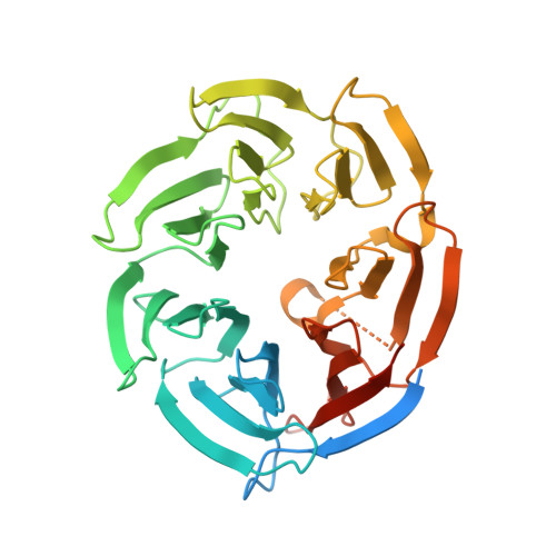
    In this study, we describe the rapid identification of potent binders for the WD40 repeat domain (WDR) of DCAF1. This was achieved by two rounds of iterative focused screening of a small set of compounds selected on the basis of internal WDR domain knowledge followed by hit expansion. Subsequent structure-based design led to nanomolar potency binders with a clear exit vector enabling DCAF1-based bifunctional degrader exploration.


  • Organizational Affiliation

    Global Discovery Chemistry, Novartis Institutes for BioMedical Research, Basel 4002, Switzerland.


Macromolecules
Find similar proteins by:  (by identity cutoff)  |  3D Structure
Entity ID: 1
MoleculeChains Sequence LengthOrganismDetailsImage
DDB1- and CUL4-associated factor 1367Homo sapiensMutation(s): 1 
Gene Names: DCAF1KIAA0800RIPVPRBP
EC: 2.7.11.1
UniProt & NIH Common Fund Data Resources
Find proteins for Q9Y4B6 (Homo sapiens)
Explore Q9Y4B6 
Go to UniProtKB:  Q9Y4B6
PHAROS:  Q9Y4B6
GTEx:  ENSG00000145041 
Entity Groups  
Sequence Clusters30% Identity50% Identity70% Identity90% Identity95% Identity100% Identity
UniProt GroupQ9Y4B6
Sequence Annotations
Expand
  • Reference Sequence
Experimental Data & Validation

Experimental Data

  • Method: X-RAY DIFFRACTION
  • Resolution: 2.20 Å
  • R-Value Free: 0.235 
  • R-Value Work: 0.197 
  • Space Group: P 61 2 2
Unit Cell:
Length ( Å )Angle ( ˚ )
a = 81.924α = 90
b = 81.924β = 90
c = 236.778γ = 120
Software Package:
Software NamePurpose
REFMACrefinement
Aimlessdata scaling
XDSdata reduction
PHASERphasing

Structure Validation

View Full Validation Report


Ligand Structure Quality Assessment 


Entry History & Funding Information

Deposition Data


Funding OrganizationLocationGrant Number
Not funded--

Revision History  (Full details and data files)

  • Version 1.0: 2023-06-14
    Type: Initial release
  • Version 1.1: 2023-08-02
    Changes: Database references