8ODZ | pdb_00008odz

Cryo-EM structure of a pre-dimerized murine IL-12 complete extracellular signaling complex (Class 1).


Experimental Data Snapshot

  • Method: ELECTRON MICROSCOPY
  • Resolution: 3.60 Å
  • Aggregation State: PARTICLE 
  • Reconstruction Method: SINGLE PARTICLE 

wwPDB Validation 3D Report Full Report

Validation slider image for 8ODZ

This is version 1.2 of the entry. See complete history

Literature

Structures of complete extracellular receptor assemblies mediated by IL-12 and IL-23.

Bloch, Y.Felix, J.Merceron, R.Provost, M.Symakani, R.A.De Backer, R.Lambert, E.Mehdipour, A.R.Savvides, S.N.

(2024) Nat Struct Mol Biol 31: 591-597

  • DOI: https://doi.org/10.1038/s41594-023-01190-6
  • Primary Citation Related Structures: 
    8C7M, 8CR5, 8CR6, 8CR8, 8ODX, 8ODZ, 8OE0, 8OE4, 8PB1, 8PPM

  • PubMed Abstract: 

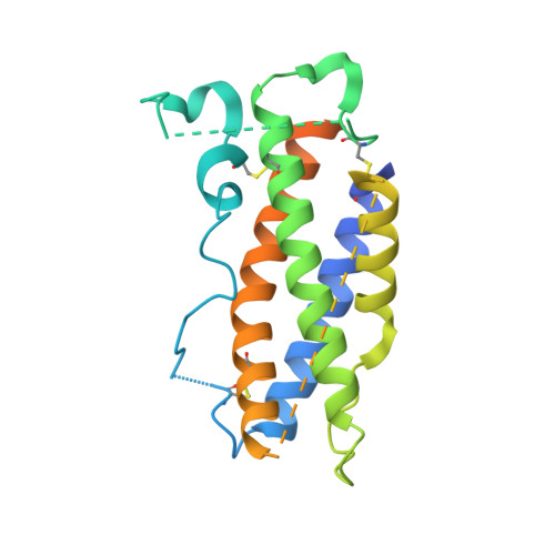
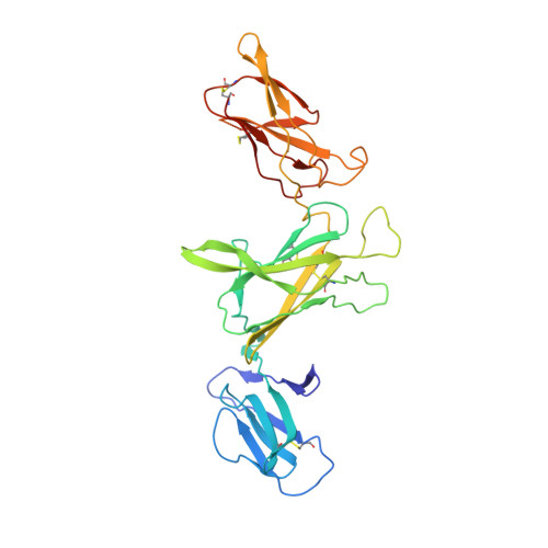
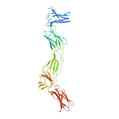
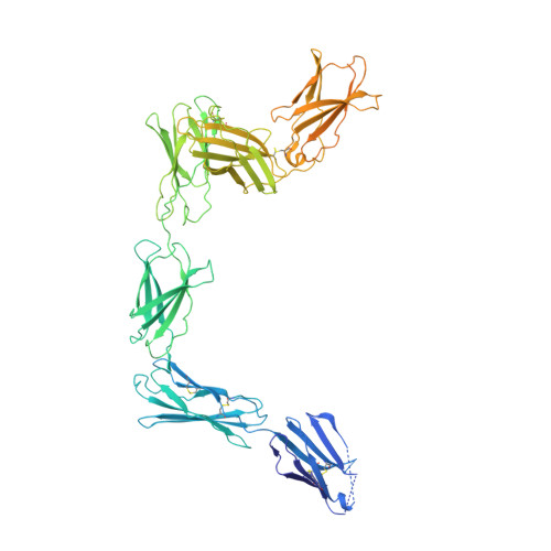
    Cell-surface receptor complexes mediated by pro-inflammatory interleukin (IL)-12 and IL-23, both validated therapeutic targets, are incompletely understood due to the lack of structural insights into their complete extracellular assemblies. Furthermore, there is a paucity of structural details describing the IL-12-receptor interaction interfaces, in contrast to IL-23-receptor complexes. Here we report structures of fully assembled mouse IL-12/human IL-23-receptor complexes comprising the complete extracellular segments of the cognate receptors determined by electron cryo-microscopy. The structures reveal key commonalities but also surprisingly diverse features. Most notably, whereas IL-12 and IL-23 both utilize a conspicuously presented aromatic residue on their α-subunit as a hotspot to interact with the N-terminal Ig domain of their high-affinity receptors, only IL-12 juxtaposes receptor domains proximal to the cell membrane. Collectively, our findings will help to complete our understanding of cytokine-mediated assemblies of tall cytokine receptors and will enable a cytokine-specific interrogation of IL-12/IL-23 signaling in physiology and disease.


  • Organizational Affiliation
    • Unit for Structural Biology, Department of Biochemistry and Microbiology, Ghent University, Ghent, Belgium.

Macromolecule Content 

  • Total Structure Weight: 213.13 kDa 
  • Atom Count: 12,100 
  • Modeled Residue Count: 1,516 
  • Deposited Residue Count: 1,885 
  • Unique protein chains: 4

Macromolecules

Find similar proteins by:|  3D Structure
Entity ID: 1
MoleculeChains  Sequence LengthOrganismDetailsImage
Interleukin-12 subunit alpha231Mus musculusMutation(s): 0 
Gene Names: Il12a
UniProt & NIH Common Fund Data Resources
Find proteins for P43431 (Mus musculus)
Explore P43431 
Go to UniProtKB:  P43431
IMPC:  MGI:96539
Entity Groups
Sequence Clusters30% Identity50% Identity70% Identity90% Identity95% Identity100% Identity
UniProt GroupP43431
Sequence Annotations
Expand
Reference Sequence
Find similar proteins by:|  3D Structure
Entity ID: 2
MoleculeChains  Sequence LengthOrganismDetailsImage
Interleukin-12 subunit beta313Mus musculusMutation(s): 0 
Gene Names: Il12b
UniProt & NIH Common Fund Data Resources
Find proteins for P43432 (Mus musculus)
Explore P43432 
Go to UniProtKB:  P43432
IMPC:  MGI:96540
Entity Groups
Sequence Clusters30% Identity50% Identity70% Identity90% Identity95% Identity100% Identity
UniProt GroupP43432
Glycosylation
Glycosylation Sites: 1Go to GlyGen: P43432-1
Sequence Annotations
Expand
Reference Sequence
Find similar proteins by:|  3D Structure
Entity ID: 3
MoleculeChains  Sequence LengthOrganismDetailsImage
Interleukin-12 receptor subunit beta-1,Death-associated protein kinase 1572Mus musculusMutation(s): 0 
Gene Names: Il12rb1Il12rbDAPK1DAPK
EC: 2.7.11.1
UniProt & NIH Common Fund Data Resources
Find proteins for P53355 (Homo sapiens)
Explore P53355 
Go to UniProtKB:  P53355
PHAROS:  P53355
GTEx:  ENSG00000196730 
Entity Groups
Sequence Clusters30% Identity50% Identity70% Identity90% Identity95% Identity100% Identity
UniProt GroupP53355
Glycosylation
Glycosylation Sites: 3Go to GlyGen: P53355-1
Sequence Annotations
Expand
Reference Sequence
Find similar proteins by:|  3D Structure
Entity ID: 4
MoleculeChains  Sequence LengthOrganismDetailsImage
Interleukin-12 receptor subunit beta-2,Calmodulin-1769Mus musculusMutation(s): 0 
Gene Names: Il12rb2CALM1CALMCAMCAM1
UniProt
Find proteins for P0DP26 (Mus musculus)
Explore P0DP26 
Go to UniProtKB:  P0DP26
Find proteins for P97378 (Mus musculus)
Explore P97378 
Go to UniProtKB:  P97378
Entity Groups
Sequence Clusters30% Identity50% Identity70% Identity90% Identity95% Identity100% Identity
UniProt GroupsP97378P0DP26
Glycosylation
Glycosylation Sites: 2Go to GlyGen: P97378-1P0DP26-1
Sequence Annotations
Expand
Reference Sequence

Oligosaccharides

Help  
Entity ID: 5
MoleculeChains Length2D Diagram GlycosylationD Interactions
alpha-D-mannopyranose-(1-3)-beta-D-mannopyranose-(1-4)-2-acetamido-2-deoxy-beta-D-glucopyranose-(1-4)-2-acetamido-2-deoxy-beta-D-glucopyranose
E
4N-Glycosylation
Glycosylation Resources
GlyTouCan: G81315DD
GlyCosmos: G81315DD
GlyGen: G81315DD

Experimental Data & Validation

Experimental Data

  • Method: ELECTRON MICROSCOPY
  • Resolution: 3.60 Å
  • Aggregation State: PARTICLE 
  • Reconstruction Method: SINGLE PARTICLE 
EM Software:
TaskSoftware PackageVersion
MODEL REFINEMENTPHENIX1.19.2-4158
RECONSTRUCTIONcryoSPARC

Structure Validation

View Full Validation Report


Entry History 

& Funding Information

Deposition Data


Funding OrganizationLocationGrant Number
Research Foundation - Flanders (FWO)Belgium12S0519N
Research Foundation - Flanders (FWO)BelgiumG0B4918N

Revision History  (Full details and data files)

  • Version 1.0: 2024-02-07
    Type: Initial release
  • Version 1.1: 2024-05-01
    Changes: Database references
  • Version 1.2: 2024-11-13
    Changes: Data collection, Structure summary