


Phosphate-Binding Protein (PstS) from Xanthomonas citri pv. citri A306
Santos, L.S., Balan, A., Guskov, A.To be published.
Experimental Data Snapshot
Starting Model: in silico
View more details
Entity ID: 1 | |||||
|---|---|---|---|---|---|
| Molecule | Chains | Sequence Length | Organism | Details | Image |
| Phosphate-binding protein PstS | 344 | Xanthomonas citri subsp. citri 306 | Mutation(s): 0 Gene Names: pstS | 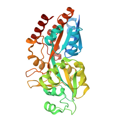 | |
| Ligands 1 Unique | |||||
|---|---|---|---|---|---|
| ID | Chains | Name / Formula / InChI Key | 2D Diagram | 3D Interactions | |
| PO4 (Subject of Investigation/LOI) Download:Ideal Coordinates CCD File | B [auth A] | PHOSPHATE ION O4 P NBIIXXVUZAFLBC-UHFFFAOYSA-K |  | ||
| Length ( Å ) | Angle ( ˚ ) |
|---|---|
| a = 43.37 | α = 90 |
| b = 73.137 | β = 90 |
| c = 99.165 | γ = 90 |
| Software Name | Purpose |
|---|---|
| BUSTER | refinement |
| XDS | data reduction |
| XSCALE | data scaling |
| BALBES | phasing |
| Funding Organization | Location | Grant Number |
|---|---|---|
| Sao Paulo Research Foundation (FAPESP) | Brazil | 2020/10171-0 |


