


Crystal structure of S-adenosylmethionine-dependent methyltransferase from Fusobacterium nucleatum
He, S.R., Bai, X.To be published.
Experimental Data Snapshot
wwPDB Validation 3D Report Full Report

Entity ID: 1 | |||||
|---|---|---|---|---|---|
| Molecule | Chains | Sequence Length | Organism | Details | Image |
| S-adenosylmethionine-dependent methyltransferase | 249 | Fusobacterium nucleatum subsp. nucleatum ATCC 25586 | Mutation(s): 0 Gene Names: FN1919 EC: 2.1.1 | 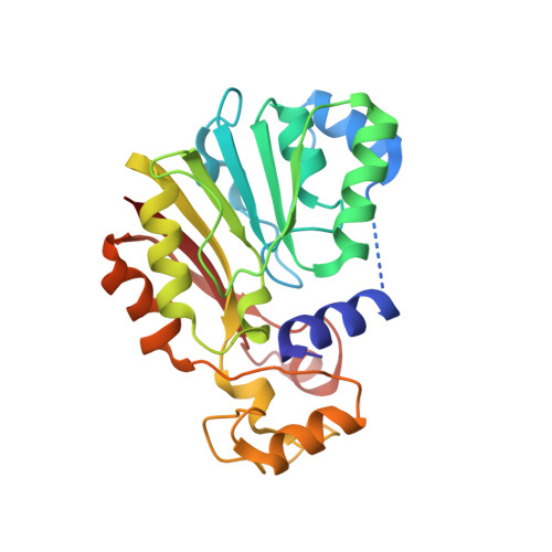 | |
UniProt | |||||
Entity Groups | |||||
| Sequence Clusters | 30% Identity50% Identity70% Identity90% Identity95% Identity100% Identity | ||||
| UniProt Group | Q8R6D8 | ||||
Sequence AnnotationsExpand | |||||
Reference Sequence | |||||
| Length ( Å ) | Angle ( ˚ ) |
|---|---|
| a = 62.981 | α = 90 |
| b = 80.629 | β = 90 |
| c = 108.49 | γ = 90 |
| Software Name | Purpose |
|---|---|
| PHENIX | refinement |
| HKL-3000 | data reduction |
| HKL-3000 | data scaling |
| PHENIX | phasing |
| Funding Organization | Location | Grant Number |
|---|---|---|
| National Research Foundation (NRF, Korea) | Korea, Republic Of | -- |


