Discovery of Novel PD-L1 Inhibitors That Induce the Dimerization, Internalization, and Degradation of PD-L1 Based on the Fragment Coupling Strategy.
Wang, K., Zhang, X., Cheng, Y., Qi, Z., Ye, K., Zhang, K., Jiang, S., Liu, Y., Xiao, Y., Wang, T.(2023) J Med Chem 66: 16807-16827
- PubMed: 38109261 Search on PubMed
- DOI: https://doi.org/10.1021/acs.jmedchem.3c01534
- Primary Citation Related Structures:
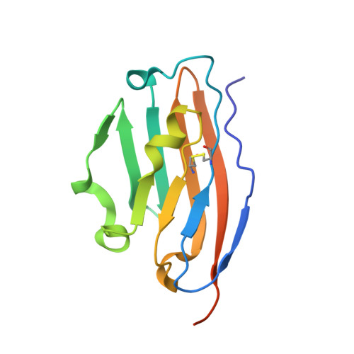
8K5N - PubMed Abstract:
Tumor cells can evade immune surveillance through overexpressing programmed cell death-ligand 1 (PD-L1) to interact with programmed cell death-1 (PD-1). Besides, tumor-intrinsic PD-L1 is involved in tumor progression without interaction with PD-1, which provides more challenges for the discovery of PD-L1 inhibitors. Herein, we report the discovery of novel PD-L1 inhibitors using the fragment coupling strategy. Among them, B9 was found to inhibit the PD-1/PD-L1 interaction with the best IC 50 value of 1.8 ± 0.7 nM. Beyond the blockade of the PD-1/PD-L1 axis, B9 promotes the dimerization, internalization, and degradation of PD-L1. Furthermore, B9 displayed high in vivo antitumor efficacy in the CT26 mouse model and activated the immune microenvironment and induced PD-L1 degradation of PD-L1 in the tumor. These results show that B9 is a promising lead PD-L1 inhibitor through the blockade of PD-1/PD-L1 interaction and functional inhibition of the PD-L1 signal pathway.
- School of Pharmacy, China Pharmaceutical University, Nanjing 210009, China.
Organizational Affiliation: