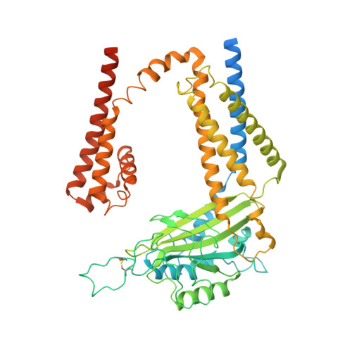
8K3S

Structure of PKD2-F604P complex


Experimental Data Snapshot

  • Method: ELECTRON MICROSCOPY
  • Resolution: 3.00 Å
  • Aggregation State: PARTICLE 
  • Reconstruction Method: SINGLE PARTICLE 

wwPDB Validation   3D Report Full Report


This is version 1.1 of the entry. See complete history


Literature

Molecular and structural basis of the dual regulation of the polycystin-2 ion channel by small-molecule ligands.

Wang, Z.Chen, M.Su, Q.Morais, T.D.C.Wang, Y.Nazginov, E.Pillai, A.R.Qian, F.Shi, Y.Yu, Y.

(2024) Proc Natl Acad Sci U S A 121: e2316230121-e2316230121

  • DOI: https://doi.org/10.1073/pnas.2316230121
  • Primary Citation of Related Structures:  
    8HK7, 8K3S

  • PubMed Abstract: 

    Mutations in the PKD2 gene, which encodes the polycystin-2 (PC2, also called TRPP2) protein, lead to autosomal dominant polycystic kidney disease (ADPKD). As a member of the transient receptor potential (TRP) channel superfamily, PC2 functions as a non-selective cation channel. The activation and regulation of the PC2 channel are largely unknown, and direct binding of small-molecule ligands to this channel has not been reported. In this work, we found that most known small-molecule agonists of the mucolipin TRP (TRPML) channels inhibit the activity of the PC2_F604P, a gain-of-function mutant of the PC2 channel. However, two of them, ML-SA1 and SF-51, have dual regulatory effects, with low concentration further activating PC2_F604P, and high concentration leading to inactivation of the channel. With two cryo-electron microscopy (cryo-EM) structures, a molecular docking model, and mutagenesis results, we identified two distinct binding sites of ML-SA1 in PC2_F604P that are responsible for activation and inactivation, respectively. These results provide structural and functional insights into how ligands regulate PC2 channel function through unusual mechanisms and may help design compounds that are more efficient and specific in regulating the PC2 channel and potentially also for ADPKD treatment.


  • Organizational Affiliation

    Department of Biological Sciences, St. John's University, Queens, NY 11375.


Macromolecules
Find similar proteins by:  (by identity cutoff)  |  3D Structure
Entity ID: 1
MoleculeChains Sequence LengthOrganismDetailsImage
Polycystin-2
A, B, C, D
571Homo sapiensMutation(s): 1 
Gene Names: PKD2TRPP2
Membrane Entity: Yes 
UniProt & NIH Common Fund Data Resources
Find proteins for Q13563 (Homo sapiens)
Explore Q13563 
Go to UniProtKB:  Q13563
GTEx:  ENSG00000118762 
Entity Groups  
Sequence Clusters30% Identity50% Identity70% Identity90% Identity95% Identity100% Identity
UniProt GroupQ13563
Glycosylation
Glycosylation Sites: 3Go to GlyGen: Q13563-1
Sequence Annotations
Expand
  • Reference Sequence
Small Molecules
Ligands 3 Unique
IDChains Name / Formula / InChI Key2D Diagram3D Interactions
PEF (Subject of Investigation/LOI)
Query on PEF

Download Ideal Coordinates CCD File 
E [auth A],
J [auth B],
N [auth C],
R [auth D]
DI-PALMITOYL-3-SN-PHOSPHATIDYLETHANOLAMINE
C37 H74 N O8 P
SLKDGVPOSSLUAI-PGUFJCEWSA-N
NAG
Query on NAG

Download Ideal Coordinates CCD File 
F [auth A]
G [auth A]
H [auth A]
K [auth B]
L [auth B]
F [auth A],
G [auth A],
H [auth A],
K [auth B],
L [auth B],
M [auth B],
O [auth C],
P [auth C],
Q [auth C],
S [auth D],
T [auth D],
U [auth D]
2-acetamido-2-deoxy-beta-D-glucopyranose
C8 H15 N O6
OVRNDRQMDRJTHS-FMDGEEDCSA-N
CA
Query on CA

Download Ideal Coordinates CCD File 
I [auth A]CALCIUM ION
Ca
BHPQYMZQTOCNFJ-UHFFFAOYSA-N
Experimental Data & Validation

Experimental Data

  • Method: ELECTRON MICROSCOPY
  • Resolution: 3.00 Å
  • Aggregation State: PARTICLE 
  • Reconstruction Method: SINGLE PARTICLE 

Structure Validation

View Full Validation Report


Entry History & Funding Information

Deposition Data


Funding OrganizationLocationGrant Number
National Natural Science Foundation of China (NSFC)China31930059

Revision History  (Full details and data files)

  • Version 1.0: 2024-04-03
    Type: Initial release
  • Version 1.1: 2024-05-22
    Changes: Database references