8K1Y | pdb_00008k1y

Crystal structure of human serum albumin and ruthenium Val complex adduct


Experimental Data Snapshot

  • Method: X-RAY DIFFRACTION
  • Resolution: 2.15 Å
  • R-Value Free: 
    0.269 (Depositor), 0.263 (DCC) 
  • R-Value Work: 
    0.225 (Depositor), 0.227 (DCC) 
  • R-Value Observed: 
    0.227 (Depositor) 

Starting Model: experimental
View more details

wwPDB Validation 3D Report Full Report

Validation slider image for 8K1Y

Ligand Structure Quality Assessment 


This is version 1.2 of the entry. See complete history

Literature

Insight into the anti-proliferation activity and photoinduced NO release of four nitrosylruthenium isomeric complexes and their HSA complex adducts.

Shi, J.Xie, L.Gong, W.Bai, H.Wang, W.Wang, A.Cao, W.Tong, H.Wang, H.

(2024) Metallomics 16

  • DOI: https://doi.org/10.1093/mtomcs/mfae005
  • Primary Citation Related Structures: 
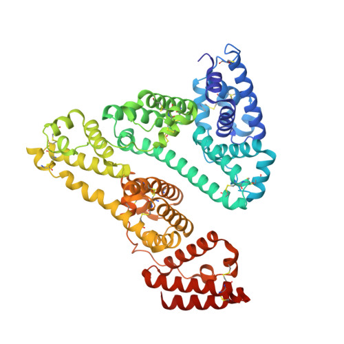
    8K1Y

  • PubMed Abstract: 

    Four Ru(II)-centered isomeric complexes [RuCl(5cqn)(Val)(NO)] (1-4) were synthesized with 5cqn (5-chloro-8-hydroxyquinoline) and chiral Val (Val = L- or D-valine) as co-ligand, and their structures were confirmed using the X-ray diffraction method. The cytotoxicity and photodynamic activity of the isomeric complexes and their human serum albumin (HSA) complex adducts were evaluated. Both the isomeric complexes and their HSA complex adducts significantly affected HeLa cell proliferation, with an IC50 value in the range of 0.3-0.5 μM. The photo-controlled release of nitric oxide (NO) in solution was confirmed using time-resolved Fourier transform infrared and electron paramagnetic resonance spectroscopy techniques. Furthermore, photoinduced NO release in living cells was observed using a selective fluorescent probe for NO. Moreover, the binding constants (Kb) of the complexes with HSA were calculated to be 0.17-1.98 × 104 M-1 and the average number of binding sites (n) was found to be close to 1, it can serve as a crucial carrier for delivering metal complexes. The crystal structure of the HSA complex adduct revealed that one [RuCl(H2O)(NO)(Val)]+ molecule binds to a pocket in domain I. This study provides insight into possible mechanism of metabolism and potential applications for nitrosylruthenium complexes.


  • Organizational Affiliation
    • Key Laboratory of Chemical Biology and Molecular Engineering of the Education Ministry and Key Laboratory of Energy Conversion and Storage Materials of Shanxi Provence, Institute of Molecular Science, Shanxi University, Taiyuan 030006, China.

Macromolecule Content 

  • Total Structure Weight: 68.89 kDa 
  • Atom Count: 4,894 
  • Modeled Residue Count: 581 
  • Deposited Residue Count: 585 
  • Unique protein chains: 1

Macromolecules

Find similar proteins by:|  3D Structure
Entity ID: 1
MoleculeChains  Sequence LengthOrganismDetailsImage
Albumin585Homo sapiensMutation(s): 0 
Gene Names: ALBGIG20GIG42PRO0903PRO1708PRO2044PRO2619PRO2675UNQ696/PRO1341
UniProt & NIH Common Fund Data Resources
Find proteins for P02768 (Homo sapiens)
Explore P02768 
Go to UniProtKB:  P02768
PHAROS:  P02768
GTEx:  ENSG00000163631 
Entity Groups
Sequence Clusters30% Identity50% Identity70% Identity90% Identity95% Identity100% Identity
UniProt GroupP02768
Sequence Annotations
Expand
Reference Sequence

Small Molecules

Ligands 7 Unique
IDChains Name / Formula / InChI Key2D Diagram3D Interactions
PLM

Query on PLM


Download:Ideal Coordinates CCD File
F [auth A]
G [auth A]
H [auth A]
I [auth A]
J [auth A]
F [auth A],
G [auth A],
H [auth A],
I [auth A],
J [auth A],
K [auth A]
PALMITIC ACID
C16 H32 O2
IPCSVZSSVZVIGE-UHFFFAOYSA-N
VAL
(Subject of Investigation/LOI)

Query on VAL


Download:Ideal Coordinates CCD File
C [auth A]VALINE
C5 H11 N O2
KZSNJWFQEVHDMF-BYPYZUCNSA-N
RU

Query on RU


Download:Ideal Coordinates CCD File
E [auth A]RUTHENIUM ION
Ru
BPEVHDGLPIIAGH-UHFFFAOYSA-N
GOL

Query on GOL


Download:Ideal Coordinates CCD File
L [auth A],
M [auth A]
GLYCEROL
C3 H8 O3
PEDCQBHIVMGVHV-UHFFFAOYSA-N
DMS

Query on DMS


Download:Ideal Coordinates CCD File
N [auth A],
O [auth A],
P [auth A],
Q [auth A]
DIMETHYL SULFOXIDE
C2 H6 O S
IAZDPXIOMUYVGZ-UHFFFAOYSA-N
CL

Query on CL


Download:Ideal Coordinates CCD File
D [auth A]CHLORIDE ION
Cl
VEXZGXHMUGYJMC-UHFFFAOYSA-M
NO

Query on NO


Download:Ideal Coordinates CCD File
B [auth A]NITRIC OXIDE
N O
ODUCDPQEXGNKDN-UHFFFAOYSA-N

Experimental Data & Validation

Experimental Data

  • Method: X-RAY DIFFRACTION
  • Resolution: 2.15 Å
  • R-Value Free:  0.269 (Depositor), 0.263 (DCC) 
  • R-Value Work:  0.225 (Depositor), 0.227 (DCC) 
  • R-Value Observed: 0.227 (Depositor) 
Space Group: C 1 2 1
Unit Cell:
Length ( Å )Angle ( ˚ )
a = 185.004α = 90
b = 38.418β = 105.18
c = 95.781γ = 90
Software Package:
Software NamePurpose
PHENIXrefinement
HKL-2000data reduction
HKL-2000data scaling
PHENIXphasing

Structure Validation

View Full Validation Report


Ligand Structure Quality Assessment 


Entry History 

& Funding Information

Deposition Data


Funding OrganizationLocationGrant Number
National Natural Science Foundation of China (NSFC)China62075118

Revision History  (Full details and data files)

  • Version 1.0: 2024-07-17
    Type: Initial release
  • Version 1.1: 2024-10-23
    Changes: Structure summary
  • Version 1.2: 2024-12-11
    Changes: Database references