8JYD | pdb_00008jyd

A genetically encoded sensor based on a bacterial DNA ligase


Experimental Data Snapshot

  • Method: X-RAY DIFFRACTION
  • Resolution: 3.00 Å
  • R-Value Free: 
    0.256 (Depositor), 0.240 (DCC) 
  • R-Value Work: 
    0.215 (Depositor), 0.230 (DCC) 
  • R-Value Observed: 
    0.217 (Depositor) 

Starting Model: experimental
View more details

wwPDB Validation   3D Report Full Report


Ligand Structure Quality Assessment 


This is version 1.1 of the entry. See complete history


Literature

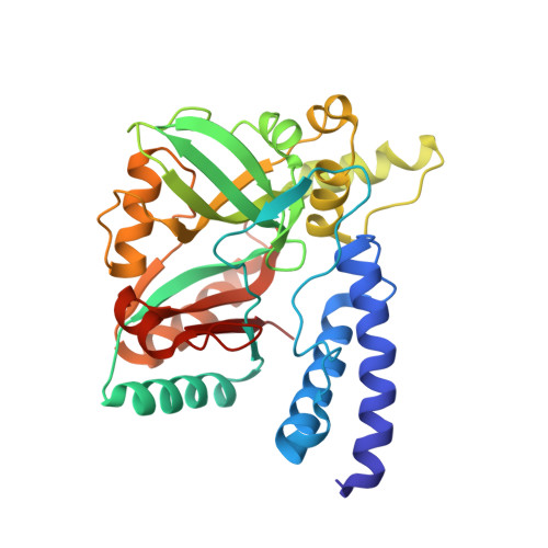
Quantitative dynamics of intracellular NMN by genetically encoded biosensor.

Chen, L.Wang, P.Huang, G.Cheng, W.Liu, K.Yu, Q.

(2025) Biosens Bioelectron 267: 116842-116842

  • DOI: https://doi.org/10.1016/j.bios.2024.116842
  • Primary Citation of Related Structures:  
    8JYD

  • PubMed Abstract: 

    Nicotinamide mononucleotide (NMN) is the direct precursor and a major booster of NAD + with increasing applications in NAD + - and aging-related pathologies. However, measuring live cell NMN dynamics was not possible, leaving key questions in NMN uptake and intracellular regulation unanswered. Here we developed genetically encoded bioluminescent and fluorescent sensors to quantify subcellular NMN in live cells by engineering specific NMN-responsive protein scaffolds fused to luciferase and fluorescent proteins. The sensor dissected the multimechanistic uptake of exogenous NMN and nicotinamide riboside (NR) in live cells and further measured the NMN levels across different subcellular compartments, as well as the perturbed NMN/NAD + ratios by external supplements. Moreover, we measured the NMN regulation by NAD(H) hydrolase Nudts and peroxisomal carrier Pxmp2 and identified Slc25a45 as a potential mitochondrial NMN regulator for its unique fingerprint on the local NMN/NAD + ratio. Collectively, the genetically encoded sensors provide a useful tool for visualizing NMN metabolism.


  • Organizational Affiliation
    • Sino-European Center of Biomedicine and Health, Institute of Biomedicine and Biotechnology, Shenzhen Institute of Advanced Technology, Chinese Academy of Sciences, 518055, Shenzhen, China.

Macromolecules
Find similar proteins by:  (by identity cutoff)  |  3D Structure
Entity ID: 1
MoleculeChains Sequence LengthOrganismDetailsImage
DNA ligase312Enterococcus faecalis V583Mutation(s): 0 
Gene Names: ligAEF_0722
EC: 6.5.1.2
UniProt
Find proteins for Q837V6 (Enterococcus faecalis (strain ATCC 700802 / V583))
Explore Q837V6 
Go to UniProtKB:  Q837V6
Entity Groups  
Sequence Clusters30% Identity50% Identity70% Identity90% Identity95% Identity100% Identity
UniProt GroupQ837V6
Sequence Annotations
Expand
  • Reference Sequence
Small Molecules
Ligands 1 Unique
IDChains Name / Formula / InChI Key2D Diagram3D Interactions
NMN (Subject of Investigation/LOI)
Query on NMN

Download Ideal Coordinates CCD File 
B [auth A]BETA-NICOTINAMIDE RIBOSE MONOPHOSPHATE
C11 H16 N2 O8 P
DAYLJWODMCOQEW-TURQNECASA-O
Experimental Data & Validation

Experimental Data

  • Method: X-RAY DIFFRACTION
  • Resolution: 3.00 Å
  • R-Value Free:  0.256 (Depositor), 0.240 (DCC) 
  • R-Value Work:  0.215 (Depositor), 0.230 (DCC) 
  • R-Value Observed: 0.217 (Depositor) 
Space Group: P 31 2 1
Unit Cell:
Length ( Å )Angle ( ˚ )
a = 92.48α = 90
b = 92.48β = 90
c = 74.29γ = 120
Software Package:
Software NamePurpose
REFMACrefinement
xia2data reduction
xia2data scaling
MoRDaphasing

Structure Validation

View Full Validation Report


Ligand Structure Quality Assessment 


Entry History & Funding Information

Deposition Data

  • Released Date: 2024-07-10 
  • Deposition Author(s): Chen, L.

Funding OrganizationLocationGrant Number
Not funded--

Revision History  (Full details and data files)

  • Version 1.0: 2024-07-10
    Type: Initial release
  • Version 1.1: 2025-01-29
    Changes: Database references, Structure summary