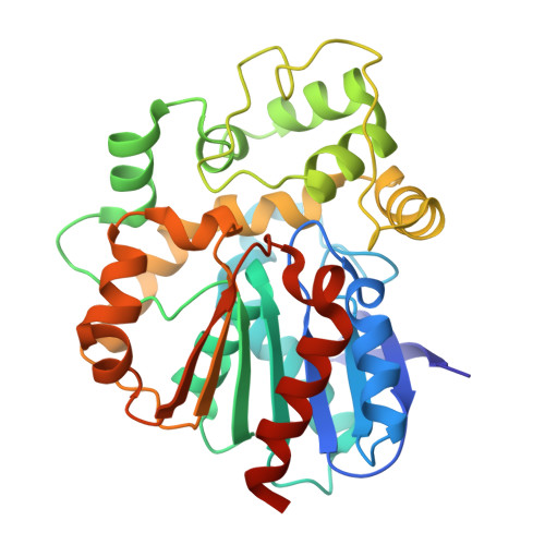
Structure of epoxide hydrolase 2 from Mangifera indica throws light on the substrate specificity determinants of plant epoxide hydrolases
Bhoite, A., Gaur, N.K., Palang, M., Kontham, R., Gupta, V., Kulkarni, K.(2024) Biochem Biophys Res Commun 733: 150444-150444
- PubMed: 39067247 Search on PubMed
- DOI: https://doi.org/10.1016/j.bbrc.2024.150444
- Primary Citation Related Structures:
8JY1 - PubMed Abstract:
Epoxide hydrolases (EHs) are a group of ubiquitous enzymes that catalyze hydrolysis of chemically reactive epoxides to yield corresponding dihydrodiols. Despite extensive studies on EHs from different clades, generic rules governing their substrate specificity determinants have remained elusive. Here, we present structural, biochemical and molecular dynamics simulation studies on MiEH2, a plant epoxide hydrolase from Mangifera indica. Comparative structure-function analysis of nine homologs of MiEH2, which include a few AlphaFold structural models, show that the two conserved tyrosines (MiEH2 Y152 and MiEH2 Y232 ) from the lid domain dissect substrate binding tunnel into two halves, forming substrate-binding-pocket one (BP1) and two (BP2). This compartmentalization offers diverse binding modes to their substrates, as exemplified by the binding of smaller aromatic substrates, such as styrene oxide (SO). Docking and molecular dynamics simulations reveal that the linear epoxy fatty acid substrates predominantly occupy BP1, while the aromatic substrates can bind to either BP1 or BP2. Furthermore, SO preferentially binds to BP2, by stacking against catalytically important histidine (MiEH2 H297 ) with the conserved lid tyrosines engaging its epoxide oxygen. Residue (MiEH2 L263 ) next to the catalytic aspartate (MiEH2 D262 ) modulates substrate binding modes. Thus, the divergent binding modes correlate with the differential affinities of the EHs for their substrates. Furthermore, long-range dynamical coupling between the lid and core domains critically influences substrate enantioselectivity in plant EHs.
- Division of Biochemical Sciences, CSIR-National Chemical Laboratory, Dr. Homi Bhabha Road, Pune- 411008, India; Academy of Scientific and Innovative Research (AcSIR), Ghaziabad- 201002, India.
Organizational Affiliation: