8JVM | pdb_00008jvm

AHS-CSF domains of phage lambda tail


Experimental Data Snapshot

  • Method: ELECTRON MICROSCOPY
  • Resolution: 3.86 Å
  • Aggregation State: PARTICLE 
  • Reconstruction Method: SINGLE PARTICLE 

wwPDB Validation 3D Report Full Report

Validation slider image for 8JVM

This is version 1.4 of the entry. See complete history

Literature

Architecture of the bacteriophage lambda tail.

Wang, C.Duan, J.Gu, Z.Ge, X.Zeng, J.Wang, J.

(2024) Structure 32: 35

  • DOI: https://doi.org/10.1016/j.str.2023.10.006
  • Primary Citation Related Structures: 
    8IYD, 8IYK, 8IYL, 8JVM, 8KGE

  • PubMed Abstract: 

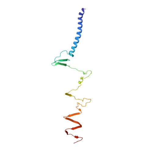
    Bacteriophage lambda has a double-stranded DNA genome and a long, flexible, non-contractile tail encoded by a contiguous block of 11 genes downstream of the head genes. The tail allows host recognition and delivery of viral DNA from the head shell to the cytoplasm of the infected cell. Here, we present a high-resolution structure of the tail complex of bacteriophage lambda determined by cryoelectron microscopy. Most component proteins of the lambda tail were determined at the atomic scale. The structure sheds light on the molecular organization of the extensively studied tail of bacteriophage lambda.


  • Organizational Affiliation
    • State Key Laboratory of Membrane Biology, Beijing Frontier Research Center for Biological Structure, School of Life Sciences, Tsinghua University, Beijing 100084, P.R. China.

Macromolecule Content 

  • Total Structure Weight: 53.2 kDa 
  • Atom Count: 3,726 
  • Modeled Residue Count: 498 
  • Deposited Residue Count: 498 
  • Unique protein chains: 1

Macromolecules

Find similar proteins by:|  3D Structure
Entity ID: 1
MoleculeChains  Sequence LengthOrganismDetailsImage
Tip attachment protein JA [auth F],
B [auth J],
C [auth Z]
166Escherichia phage LambdaMutation(s): 0 
Gene Names: Jlambdap21
UniProt
Find proteins for P03749 (Escherichia phage lambda)
Explore P03749 
Go to UniProtKB:  P03749
Entity Groups
Sequence Clusters30% Identity50% Identity70% Identity90% Identity95% Identity100% Identity
UniProt GroupP03749
Sequence Annotations
Expand
Reference Sequence

Experimental Data & Validation

Experimental Data

  • Method: ELECTRON MICROSCOPY
  • Resolution: 3.86 Å
  • Aggregation State: PARTICLE 
  • Reconstruction Method: SINGLE PARTICLE 
EM Software:
TaskSoftware PackageVersion
MODEL REFINEMENTPHENIX

Structure Validation

View Full Validation Report


Entry History 

& Funding Information

Deposition Data

  • Released Date: 2023-10-18 
  • Deposition Author(s): Wang, J.

Funding OrganizationLocationGrant Number
National Natural Science Foundation of China (NSFC)China32171190

Revision History  (Full details and data files)

  • Version 1.0: 2023-10-18
    Type: Initial release
  • Version 1.1: 2023-11-08
    Changes: Database references
  • Version 1.2: 2023-11-15
    Changes: Database references
  • Version 1.3: 2024-01-24
    Changes: Database references
  • Version 1.4: 2025-07-23
    Changes: Data collection, Structure summary