8JRL | pdb_00008jrl

Crystal structure of P450 TleB with an indole alkaloid


Experimental Data Snapshot

  • Method: X-RAY DIFFRACTION
  • Resolution: 2.07 Å
  • R-Value Free: 
    0.208 (Depositor), 0.211 (DCC) 
  • R-Value Work: 
    0.167 (Depositor), 0.168 (DCC) 
  • R-Value Observed: 
    0.169 (Depositor) 

Starting Model: experimental
View more details

wwPDB Validation 3D Report Full Report

Validation slider image for 8JRL

Ligand Structure Quality Assessment 


This is version 1.0 of the entry. See complete history

Literature

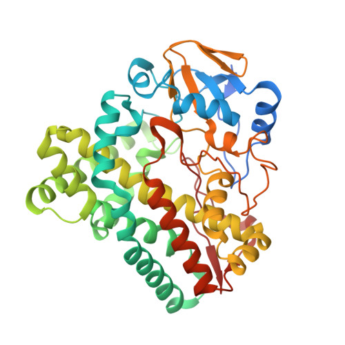
Regulation of P450 TleB catalytic flow for the synthesis of sulfur-containing indole alkaloids by substrate structure-directed strategy. and protein engineering.

Ge, X.Y.Long, Y.Wang, J.Gu, B.Yang, Z.X.Feng, Y.Y.Zheng, S.Li, Y.Y.Yan, W.P.Song, H.

(2023) Sci China B Chem 66: 3232-3241

Macromolecule Content 

  • Total Structure Weight: 47.96 kDa 
  • Atom Count: 3,410 
  • Modeled Residue Count: 392 
  • Deposited Residue Count: 418 
  • Unique protein chains: 1

Macromolecules

Find similar proteins by:|  3D Structure
Entity ID: 1
MoleculeChains  Sequence LengthOrganismDetailsImage
Cytochrome P-450418Streptomyces blastmyceticusMutation(s): 0 
Gene Names: tleB
UniProt
Find proteins for A0A077KEB8 (Streptomyces blastmyceticus)
Explore A0A077KEB8 
Go to UniProtKB:  A0A077KEB8
Entity Groups
Sequence Clusters30% Identity50% Identity70% Identity90% Identity95% Identity100% Identity
UniProt GroupA0A077KEB8
Sequence Annotations
Expand
Reference Sequence

Small Molecules

Ligands 2 Unique
IDChains Name / Formula / InChI Key2D Diagram3D Interactions
HEM
(Subject of Investigation/LOI)

Query on HEM


Download:Ideal Coordinates CCD File
B [auth A]PROTOPORPHYRIN IX CONTAINING FE
C34 H32 Fe N4 O4
KABFMIBPWCXCRK-RGGAHWMASA-L
V3X
(Subject of Investigation/LOI)

Query on V3X


Download:Ideal Coordinates CCD File
C [auth A](2~{S})-~{N}-[(2~{S})-1-(4-fluoranyl-1~{H}-indol-3-yl)-3-oxidanyl-propan-2-yl]-3-methyl-2-sulfanyl-butanamide
C16 H21 F N2 O2 S
KKPTZRIROSBPNX-NHYWBVRUSA-N

Experimental Data & Validation

Experimental Data

  • Method: X-RAY DIFFRACTION
  • Resolution: 2.07 Å
  • R-Value Free:  0.208 (Depositor), 0.211 (DCC) 
  • R-Value Work:  0.167 (Depositor), 0.168 (DCC) 
  • R-Value Observed: 0.169 (Depositor) 
Space Group: P 21 21 21
Unit Cell:
Length ( Å )Angle ( ˚ )
a = 40.035α = 90
b = 85.587β = 90
c = 113.755γ = 90
Software Package:
Software NamePurpose
PHENIXrefinement
XDSdata reduction
XDSdata scaling
PHASERphasing

Structure Validation

View Full Validation Report


Ligand Structure Quality Assessment 


Entry History 

& Funding Information

Deposition Data


Funding OrganizationLocationGrant Number
Science and Technology Commission of ShanghaiChina21ZR1433700

Revision History  (Full details and data files)

  • Version 1.0: 2024-04-17
    Type: Initial release