Download Mendeley
A conserved polar residue plays a critical role in mismatch detection in A-family DNA polymerases.
Clement, P.C., Sapam, T., Nair, D.T.(2024) Int J Biol Macromol 269: 131965-131965
- PubMed: 38697428 Search on PubMed
- DOI: https://doi.org/10.1016/j.ijbiomac.2024.131965
- Primary Citation Related Structures:
8JRB - PubMed Abstract:
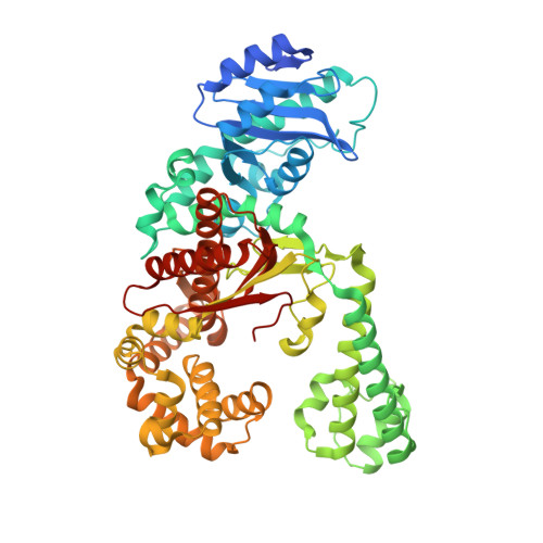
In A-family DNA polymerases (dPols), a functional 3'-5' exonuclease activity is known to proofread newly synthesized DNA. The identification of a mismatch in substrate DNA leads to transfer of the primer strand from the polymerase active site to the exonuclease active site. To shed more light regarding the mechanism responsible for the detection of mismatches, we have utilized DNA polymerase 1 from Aquifex pyrophilus (ApPol1). The enzyme synthesized DNA with high fidelity and exhibited maximal exonuclease activity with DNA substrates bearing mismatches at the -2 and - 3 positions. The crystal structure of apo-ApPol1 was utilized to generate a computational model of the functional ternary complex of this enzyme. The analysis of the model showed that N332 forms interactions with minor groove atoms of the base pairs at the -2 and - 3 positions. The majority of known A-family dPols show the presence of Asn at a position equivalent to N332. The N332L mutation led to a decrease in the exonuclease activity for representative purine-pyrimidine, and pyrimidine-pyrimidine mismatches at -2 and - 3 positions, respectively. Overall, our findings suggest that conserved polar residues located towards the minor groove may facilitate the detection of position-specific mismatches to enhance the fidelity of DNA synthesis.
- Regional Centre for Biotechnology, NCR Biotech Science Cluster, 3rd Milestone, Faridabad-Gurgaon Expressway, Faridabad 121 001, Haryana (NCR Delhi), India.
Organizational Affiliation: