8JP3 | pdb_00008jp3

FCP trimer in diatom Thalassiosira pseudonana


Experimental Data Snapshot

  • Method: ELECTRON MICROSCOPY
  • Resolution: 2.73 Å
  • Aggregation State: PARTICLE 
  • Reconstruction Method: SINGLE PARTICLE 

wwPDB Validation   3D Report Full Report


This is version 1.2 of the entry. See complete history


Literature

Structural and spectroscopic insights into fucoxanthin chlorophyll a/c-binding proteins of diatoms in diverse oligomeric states.

Zhou, C.Feng, Y.Li, Z.Shen, L.Li, X.Wang, Y.Han, G.Kuang, T.Liu, C.Shen, J.R.Wang, W.

(2024) Plant Commun 5: 101041-101041

  • DOI: https://doi.org/10.1016/j.xplc.2024.101041
  • Primary Citation of Related Structures:  
    8JP3, 8WCK, 8WCL

  • PubMed Abstract: 

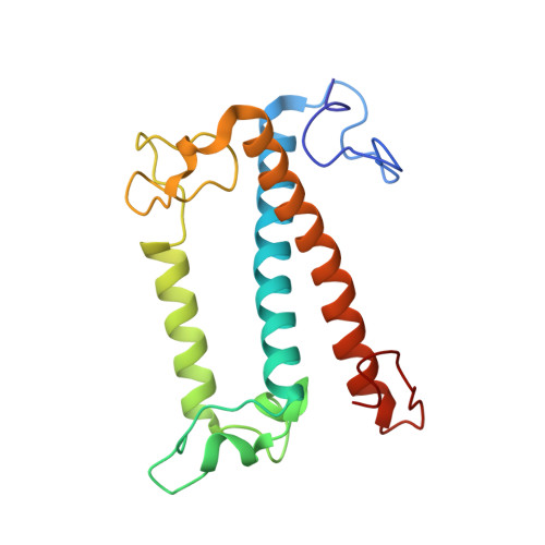
    Diatoms, a group of prevalent marine algae, significantly contribute to global primary productivity. Their substantial biomass is linked to enhanced absorption of blue-green light underwater, facilitated by fucoxanthin chlorophyll a/c-binding proteins (FCPs), exhibiting oligomeric diversity across diatom species. Utilizing mild CN-PAGE analysis on solubilized thylakoid membranes, we displayed monomeric, dimeric, trimeric, tetrameric and pentameric FCPs in diatoms. Mass spectrometry analysis revealed each oligomeric FCP has specific protein compositions, constituting a large Lhcf family of FCP antennas. In addition, we resolved the structures of Thalassiosira pseudonana FCP (Tp-FCP) homotrimer and Chaetoceros gracilis FCP (Cg-FCP) pentamer by cryo-electron microscopy at 2.73 Å and 2.65 Å resolutions, respectively. The distinct pigment composition and organization in various oligomeric FCPs change their blue-green light-harvesting, excitation energy transfer pathways. In comparison to dimeric and trimeric FCPs, Cg-FCP tetramer and Cg-FCP pentamer exhibit stronger absorption by Chls c, red-shifted and broader Chl a fluorescence emission, as well as more robust circular dichroism signals originating from Chl a-carotenoid dimers. These spectroscopic characteristics indicate that Chl a molecules in Cg-FCP tetramer and Cg-FCP pentamer are more heterogeneous than in both dimers and Tp-FCP trimer. The structural and spectroscopic insights provided by this study contribute to a better understanding of the mechanisms that empower diatoms to adapt to fluctuating light environments.


  • Organizational Affiliation
    • Photosynthesis Research Center, Key Laboratory of Photobiology, Institute of Botany, Chinese Academy of Sciences, Beijing 100093, P. R. China; University of Chinese Academy of Sciences, Beijing 100049, P. R. China.

Macromolecules
Find similar proteins by:  (by identity cutoff)  |  3D Structure
Entity ID: 1
MoleculeChains Sequence LengthOrganismDetailsImage
Fucoxanthin chlorophyll a/c protein 8
A, B, C
180Thalassiosira pseudonana CCMP1335Mutation(s): 0 
Membrane Entity: Yes 
UniProt
Find proteins for B8BS67 (Thalassiosira pseudonana)
Explore B8BS67 
Go to UniProtKB:  B8BS67
Entity Groups  
Sequence Clusters30% Identity50% Identity70% Identity90% Identity95% Identity100% Identity
UniProt GroupB8BS67
Sequence Annotations
Expand
  • Reference Sequence
Small Molecules
Ligands 5 Unique
IDChains Name / Formula / InChI Key2D Diagram3D Interactions
CLA (Subject of Investigation/LOI)
Query on CLA

Download Ideal Coordinates CCD File 
BB [auth C]
CB [auth C]
DA [auth B]
EA [auth B]
EB [auth C]
BB [auth C],
CB [auth C],
DA [auth B],
EA [auth B],
EB [auth C],
FB [auth C],
GA [auth B],
GB [auth C],
IA [auth B],
JA [auth B],
K [auth A],
L [auth A],
LA [auth B],
MA [auth B],
N [auth A],
OA [auth C],
P [auth A],
Q [auth A],
S [auth A],
T [auth A],
U [auth A],
WA [auth C],
XA [auth C],
ZA [auth C]
CHLOROPHYLL A
C55 H72 Mg N4 O5
ATNHDLDRLWWWCB-AENOIHSZSA-M
LMG (Subject of Investigation/LOI)
Query on LMG

Download Ideal Coordinates CCD File 
HB [auth C],
NA [auth B],
V [auth A]
1,2-DISTEAROYL-MONOGALACTOSYL-DIGLYCERIDE
C45 H86 O10
DCLTVZLYPPIIID-CVELTQQQSA-N
A86 (Subject of Investigation/LOI)
Query on A86

Download Ideal Coordinates CCD File 
AA [auth B]
BA [auth B]
CA [auth B]
D [auth A]
E [auth A]
AA [auth B],
BA [auth B],
CA [auth B],
D [auth A],
E [auth A],
F [auth A],
G [auth A],
H [auth A],
I [auth A],
J [auth A],
PA [auth C],
QA [auth C],
RA [auth C],
SA [auth C],
TA [auth C],
UA [auth C],
VA [auth C],
W [auth B],
X [auth B],
Y [auth B],
Z [auth B]
(3S,3'S,5R,5'R,6S,6'R,8'R)-3,5'-dihydroxy-8-oxo-6',7'-didehydro-5,5',6,6',7,8-hexahydro-5,6-epoxy-beta,beta-caroten-3'- yl acetate
C42 H58 O6
SJWWTRQNNRNTPU-XJUZQKKNSA-N
KC1 (Subject of Investigation/LOI)
Query on KC1

Download Ideal Coordinates CCD File 
DB [auth C],
KA [auth B],
R [auth A]
Chlorophyll c1
C35 H30 Mg N4 O5
DGNIJJSSARBJSH-QIEHNWLWSA-L
KC2 (Subject of Investigation/LOI)
Query on KC2

Download Ideal Coordinates CCD File 
AB [auth C]
FA [auth B]
HA [auth B]
M [auth A]
O [auth A]
AB [auth C],
FA [auth B],
HA [auth B],
M [auth A],
O [auth A],
YA [auth C]
Chlorophyll c2
C35 H28 Mg N4 O5
QDRBYWCRXZZVLY-QIEHNWLWSA-L
Experimental Data & Validation

Experimental Data

  • Method: ELECTRON MICROSCOPY
  • Resolution: 2.73 Å
  • Aggregation State: PARTICLE 
  • Reconstruction Method: SINGLE PARTICLE 
EM Software:
TaskSoftware PackageVersion
MODEL REFINEMENTPHENIX1.19.2_4158:

Structure Validation

View Full Validation Report


Entry History & Funding Information

Deposition Data


Funding OrganizationLocationGrant Number
Chinese Academy of SciencesChina31970260

Revision History  (Full details and data files)

  • Version 1.0: 2024-06-12
    Type: Initial release
  • Version 1.1: 2024-07-31
    Changes: Data collection, Database references
  • Version 1.2: 2024-11-27
    Changes: Data collection, Database references, Structure summary