8JP2

Crystal structure of AKR1C1 in complex with DFV


Experimental Data Snapshot

  • Method: X-RAY DIFFRACTION
  • Resolution: 1.80 Å
  • R-Value Free: 0.249 
  • R-Value Work: 0.197 
  • R-Value Observed: 0.199 

wwPDB Validation   3D Report Full Report


Ligand Structure Quality Assessment 


This is version 1.0 of the entry. See complete history


Literature

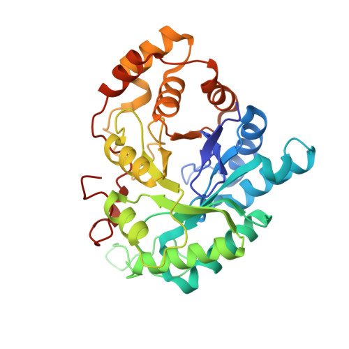
Inhibition of AKR1Cs by liquiritigenin and the structural basis.

Liu, H.Yao, Z.Sun, M.Zhang, C.Huang, Y.Y.Luo, H.B.Wu, D.Zheng, X.

(2023) Chem Biol Interact 385: 110654-110654

  • DOI: https://doi.org/10.1016/j.cbi.2023.110654
  • Primary Citation of Related Structures:  
    8JP1, 8JP2

  • PubMed Abstract: 

    In vivo and in vitro studies have confirmed that liquiritigenin (LQ), the primary active component of licorice, acts as an antitumor agent. However, how LQ diminishes or inhibits tumor growth is not fully understood. Here, we report the enzymatic inhibition of LQ and six other flavanone analogues towards AKR1Cs (AKR1C1, AKR1C2 and AKR1C3), which are involved in prostate cancer, breast cancer, and resistance of anticancer drugs. Crystallographic studies revealed AKR1C3 inhibition of LQ is related to its complementarity with the active site and the hydrogen bonds net in the catalytic site formed through C 7 -OH, aided by its nonplanar and compact structure due to the saturation of the C 2 C 3 double bond. Comparison of the LQ conformations in the structures of AKR1C1 and AKR1C3 revealed the induced-fit conformation changes, which explains the lack of isoform selectivity of LQ. Our findings will be helpful for better understanding the antitumor effects of LQ on hormonally dependent cancers and the rational design of selective AKR1Cs inhibitors.


  • Organizational Affiliation

    Key Laboratory of Molecular Target & Clinical Pharmacology and the State Key Laboratory of Respiratory Disease, School of Pharmaceutical Sciences & the Fifth Affiliated Hospital, Guangzhou Medical University, Guangzhou, 511436, China.


Macromolecules
Find similar proteins by:  (by identity cutoff)  |  3D Structure
Entity ID: 1
MoleculeChains Sequence LengthOrganismDetailsImage
Aldo-keto reductase family 1 member C1323Homo sapiensMutation(s): 0 
Gene Names: AKR1C1DDHDDH1
EC: 1.1.1 (PDB Primary Data), 1.1.1.112 (PDB Primary Data), 1.1.1.209 (PDB Primary Data), 1.1.1.210 (PDB Primary Data), 1.1.1.357 (PDB Primary Data), 1.1.1.51 (PDB Primary Data), 1.1.1.53 (PDB Primary Data), 1.1.1.62 (PDB Primary Data), 1.3.1.20 (PDB Primary Data), 1.1.1.149 (PDB Primary Data)
UniProt & NIH Common Fund Data Resources
Find proteins for Q04828 (Homo sapiens)
Explore Q04828 
Go to UniProtKB:  Q04828
PHAROS:  Q04828
GTEx:  ENSG00000187134 
Entity Groups  
Sequence Clusters30% Identity50% Identity70% Identity90% Identity95% Identity100% Identity
UniProt GroupQ04828
Sequence Annotations
Expand
  • Reference Sequence
Small Molecules
Ligands 2 Unique
IDChains Name / Formula / InChI Key2D Diagram3D Interactions
NAP (Subject of Investigation/LOI)
Query on NAP

Download Ideal Coordinates CCD File 
B [auth A]NADP NICOTINAMIDE-ADENINE-DINUCLEOTIDE PHOSPHATE
C21 H28 N7 O17 P3
XJLXINKUBYWONI-NNYOXOHSSA-N
DFV (Subject of Investigation/LOI)
Query on DFV

Download Ideal Coordinates CCD File 
C [auth A]7-HYDROXY-2-(4-HYDROXY-PHENYL)-CHROMAN-4-ONE
C15 H12 O4
FURUXTVZLHCCNA-AWEZNQCLSA-N
Experimental Data & Validation

Experimental Data

  • Method: X-RAY DIFFRACTION
  • Resolution: 1.80 Å
  • R-Value Free: 0.249 
  • R-Value Work: 0.197 
  • R-Value Observed: 0.199 
  • Space Group: P 1 21 1
Unit Cell:
Length ( Å )Angle ( ˚ )
a = 39.462α = 90
b = 83.837β = 90
c = 49.08γ = 90
Software Package:
Software NamePurpose
PHENIXrefinement
CrysalisProdata reduction
SCALAdata scaling

Structure Validation

View Full Validation Report


Ligand Structure Quality Assessment 


Entry History & Funding Information

Deposition Data


Funding OrganizationLocationGrant Number
Other government2022A1515010040

Revision History  (Full details and data files)

  • Version 1.0: 2024-04-24
    Type: Initial release