8JHC | pdb_00008jhc

FZD3 in inactive state


Experimental Data Snapshot

  • Method: ELECTRON MICROSCOPY
  • Resolution: 3.30 Å
  • Aggregation State: PARTICLE 
  • Reconstruction Method: SINGLE PARTICLE 

wwPDB Validation 3D Report Full Report

Validation slider image for 8JHC

This is version 1.1 of the entry. See complete history

Literature

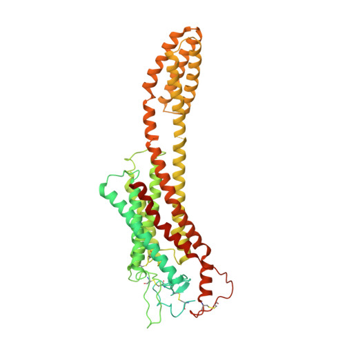
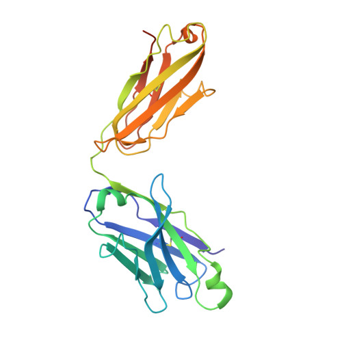
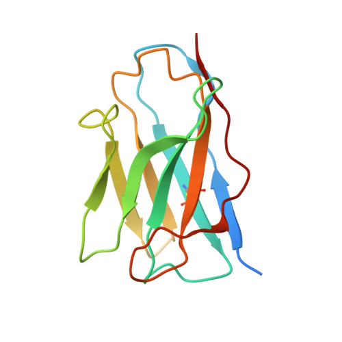
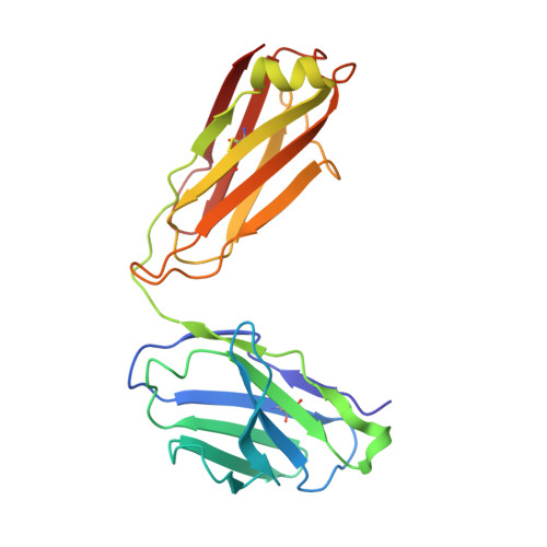
A framework for Frizzled-G protein coupling and implications to the PCP signaling pathways.

Zhang, Z.Lin, X.Wei, L.Wu, Y.Xu, L.Wu, L.Wei, X.Zhao, S.Zhu, X.Xu, F.

(2024) Cell Discov 10: 3-3

  • DOI: https://doi.org/10.1038/s41421-023-00627-y
  • Primary Citation Related Structures: 
    8J9N, 8J9O, 8JH7, 8JHB, 8JHC, 8JHI

  • PubMed Abstract: 

    The ten Frizzled receptors (FZDs) are essential in Wnt signaling and play important roles in embryonic development and tumorigenesis. Among these, FZD6 is closely associated with lens development. Understanding FZD activation mechanism is key to unlock these emerging targets. Here we present the cryo-EM structures of FZD6 and FZD3 which are known to relay non-canonical planar cell polarity (PCP) signaling pathways as well as FZD1 in their G protein-coupled states and in the apo inactive states, respectively. Comparison of the three inactive/active pairs unveiled a shared activation framework among all ten FZDs. Mutagenesis along with imaging and functional analysis on the human lens epithelial tissues suggested potential crosstalk between the G-protein coupling of FZD6 and the PCP signaling pathways. Together, this study provides an integrated understanding of FZD structure and function, and lays the foundation for developing therapeutic modulators to activate or inhibit FZD signaling for a range of disorders including cancers and cataracts.


  • Organizational Affiliation
    • iHuman Institute, ShanghaiTech University, Shanghai, China.

Macromolecule Content 

  • Total Structure Weight: 137.94 kDa 
  • Atom Count: 7,563 
  • Modeled Residue Count: 986 
  • Deposited Residue Count: 1,229 
  • Unique protein chains: 4

Macromolecules

Find similar proteins by:|  3D Structure
Entity ID: 1
MoleculeChains  Sequence LengthOrganismDetailsImage
Frizzled-3,Soluble cytochrome b562A [auth R]643Homo sapiensEscherichia coliMutation(s): 0 
Membrane Entity: Yes 
UniProt & NIH Common Fund Data Resources
Find proteins for P0ABE7 (Escherichia coli)
Explore P0ABE7 
Go to UniProtKB:  P0ABE7
Find proteins for Q9NPG1 (Homo sapiens)
Explore Q9NPG1 
Go to UniProtKB:  Q9NPG1
PHAROS:  Q9NPG1
GTEx:  ENSG00000104290 
Entity Groups
Sequence Clusters30% Identity50% Identity70% Identity90% Identity95% Identity100% Identity
UniProt GroupsP0ABE7Q9NPG1
Sequence Annotations
Expand
Reference Sequence
Find similar proteins by:|  3D Structure
Entity ID: 2
MoleculeChains  Sequence LengthOrganismDetailsImage
anti-BRIL Fab Heavy chainB [auth H]240Homo sapiensMutation(s): 0 
Find similar proteins by:|  3D Structure
Entity ID: 3
MoleculeChains  Sequence LengthOrganismDetailsImage
anti-Fab NanobodyC [auth K]134synthetic constructMutation(s): 0 
Find similar proteins by:|  3D Structure
Entity ID: 4
MoleculeChains  Sequence LengthOrganismDetailsImage
anti-BRIL Fab Light chainD [auth L]212Homo sapiensMutation(s): 0 

Experimental Data & Validation

Experimental Data

  • Method: ELECTRON MICROSCOPY
  • Resolution: 3.30 Å
  • Aggregation State: PARTICLE 
  • Reconstruction Method: SINGLE PARTICLE 

Structure Validation

View Full Validation Report


Entry History 

& Funding Information

Deposition Data


Funding OrganizationLocationGrant Number
Not funded--

Revision History  (Full details and data files)

  • Version 1.0: 2024-01-17
    Type: Initial release
  • Version 1.1: 2024-11-13
    Changes: Data collection, Structure summary