Multistep conformational changes leading to the gate opening of light-driven sodium pump rhodopsin.
Sato, Y., Hashimoto, T., Kato, K., Okamura, A., Hasegawa, K., Shinone, T., Tanaka, Y., Tanaka, Y., Tsukazaki, T., Tsukamoto, T., Demura, M., Yao, M., Kikukawa, T.(2023) J Biological Chem 299: 105393-105393
- PubMed: 37890784 Search on PubMedSearch on PubMed Central
- DOI: https://doi.org/10.1016/j.jbc.2023.105393
- Primary Citation Related Structures:
8JH0 - PubMed Abstract:
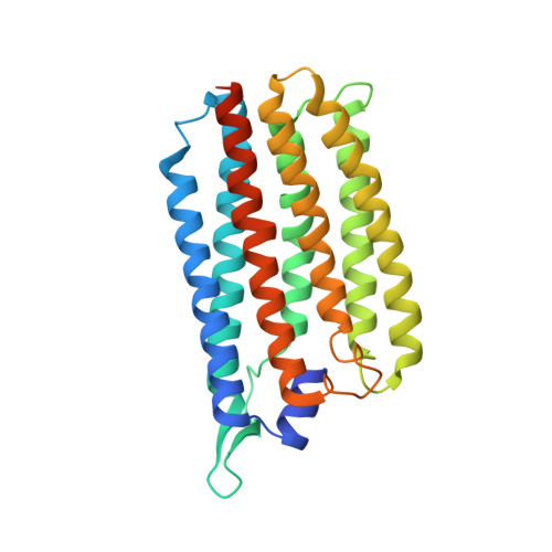
Membrane transport proteins require a gating mechanism that opens and closes the substrate transport pathway to carry out unidirectional transport. The "gating" involves large conformational changes and is achieved via multistep reactions. However, these elementary steps have not been clarified for most transporters due to the difficulty of detecting the individual steps. Here, we propose these steps for the gate opening of the bacterial Na + pump rhodopsin, which outwardly pumps Na + upon illumination. We herein solved an asymmetric dimer structure of Na + pump rhodopsin from the bacterium Indibacter alkaliphilus. In one protomer, the Arg108 sidechain is oriented toward the protein center and appears to block a Na + release pathway to the extracellular (EC) medium. In the other protomer, however, this sidechain swings to the EC side and then opens the release pathway. Assuming that the latter protomer mimics the Na + -releasing intermediate, we examined the mechanism for the swing motion of the Arg108 sidechain. On the EC surface of the first protomer, there is a characteristic cluster consisting of Glu10, Glu159, and Arg242 residues connecting three helices. In contrast, this cluster is disrupted in the second protomer. Our experimental results suggested that this disruption is a key process. The cluster disruption induces the outward movement of the Glu159-Arg242 pair and simultaneously rotates the seventh transmembrane helix. This rotation resultantly opens a space for the swing motion of the Arg108 sidechain. Thus, cluster disruption might occur during the photoreaction and then trigger sequential conformation changes leading to the gate-open state.
- Graduate School of Life Science, Hokkaido University, Sapporo, Japan.
Organizational Affiliation: