A high-resolution structural characterization and physicochemical study of how a peptoid binds to an oncoprotein MDM2.
Yokomine, M., Morimoto, J., Fukuda, Y., Ueda, T., Takeuchi, K., Umezawa, K., Ago, H., Matsuura, H., Ueno, G., Senoo, A., Nagatoishi, S., Tsumoto, K., Sando, S.(2024) Chem Sci 15: 7051-7060
- PubMed: 38756815 Search on PubMedSearch on PubMed Central
- DOI: https://doi.org/10.1039/d4sc01540a
- Primary Citation Related Structures:
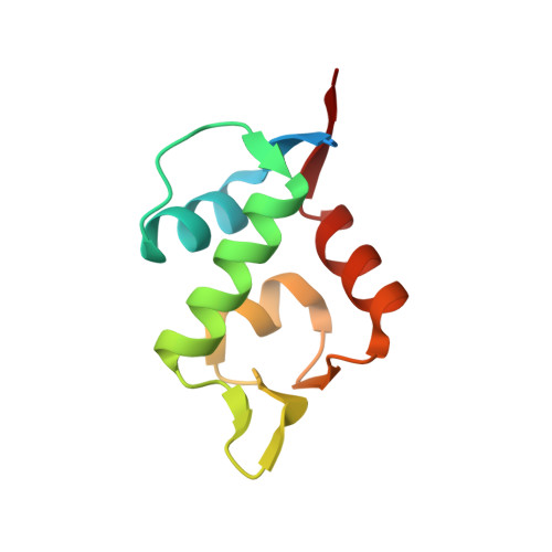
8J81 - PubMed Abstract:
Peptoids are a promising drug modality targeting disease-related proteins, but how a peptoid engages in protein binding is poorly understood. This is primarily due to a lack of high-resolution peptoid-protein complex structures and systematic physicochemical studies. Here, we present the first crystal structure of a peptoid bound to a protein, providing high-resolution structural information about how a peptoid binds to a protein. We previously reported a rigid peptoid, oligo( N -substituted alanine) (oligo-NSA), and developed an oligo-NSA-type peptoid that binds to MDM2. X-ray crystallographic analysis of the peptoid bound to MDM2 showed that the peptoid recognizes the MDM2 surface predominantly through the interaction of the N -substituents, while the main chain acts as a scaffold. Additionally, conformational, thermodynamic, and kinetic analysis of the peptoid and its derivatives with a less rigid main chain revealed that rigidification of the peptoid main chain contributes to improving the protein binding affinity. This improvement is thermodynamically attributed to an increased magnitude of the binding enthalpy change, and kinetically to an increased association rate and decreased dissociation rate. This study provides invaluable insights into the design of protein-targeting peptoids.
- Department of Chemistry and Biotechnology, Graduate School of Engineering, The University of Tokyo 7-3-1 Hongo Bunkyo-ku Tokyo 113-8656 Japan jmorimoto@chembio.t.u-tokyo.ac.jp ssando@chembio.t.u-tokyo.ac.jp.
Organizational Affiliation: